 Movie
Movie Controller


+ Open data

- Basic information
| Entry | Database: PDB / ID: 8ozw | ||||||
|---|---|---|---|---|---|---|---|
| Title | Imine Reductase from Ajellomyces dermatitidis in complex NADPH4 | ||||||
 Components Components | Oxidoreductase | ||||||
 Keywords Keywords | OXIDOREDUCTASE / Imine reductase / Amine / Dehydrogenase / NADP | ||||||
| Function / homology | 3-hydroxyisobutyrate dehydrogenase-related / 6-phosphogluconate dehydrogenase, NADP-binding / NAD binding domain of 6-phosphogluconate dehydrogenase / 6-phosphogluconate dehydrogenase, domain 2 / NADP binding / oxidoreductase activity / NAD(P)-binding domain superfamily / Chem-TXP / Oxidoreductase Function and homology information Function and homology information | ||||||
| Biological species |  Blastomyces dermatitidis (fungus) Blastomyces dermatitidis (fungus) | ||||||
| Method |  X-RAY DIFFRACTION / SYNCHROTRON / MOLECULAR REPLACEMENT / Resolution: 2.01 Å X-RAY DIFFRACTION / SYNCHROTRON / MOLECULAR REPLACEMENT / Resolution: 2.01 Å | ||||||
 Authors Authors | Sharma, M. / Grogan, G. | ||||||
| Funding support |  United Kingdom, 1items United Kingdom, 1items
| ||||||
 Citation Citation | Journal: Acta Crystallogr.,Sect.F / Year: 2023 Title: Structure of the imine reductase from Ajellomyces dermatitidis in three crystal forms. Authors: Sharma, M. / Cuetos, A. / Willliams, A. / Gonzalez-Martinez, D. / Grogan, G. | ||||||
| History |
|
- Structure visualization
| Structure viewer | Molecule: MolmilJmol/JSmol |
|---|
- Downloads & links
-Download
| PDBx/mmCIF format | 8ozw.cif.gz | 125.9 KB | Display | PDBx/mmCIF format |
|---|---|---|---|---|
| PDB format | pdb8ozw.ent.gz | 94.3 KB | Display | PDB format |
| PDBx/mmJSON format | 8ozw.json.gz | Tree view | PDBx/mmJSON format | |
| Others |  Other downloads Other downloads |
-Validation report
| Arichive directory | https://data.pdbj.org/pub/pdb/validation_reports/oz/8ozwftp://data.pdbj.org/pub/pdb/validation_reports/oz/8ozw | HTTPS FTP |
|---|
-Related structure data


-Links
 PDBj
PDBj
- Assembly
| Deposited unit | 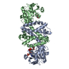
| ||||||||
|---|---|---|---|---|---|---|---|---|---|
| 1 |
| ||||||||
| Unit cell |
|
-Components
| #1: Protein | Mass: 30128.572 Da / Num. of mol.: 2 Source method: isolated from a genetically manipulated source Source: (gene. exp.) Blastomyces dermatitidis (fungus) / Gene: BDCG_07742 / Production host:  #2: Chemical | ChemComp-TXP / | 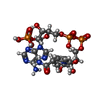  Mass: 747.437 Da / Num. of mol.: 1 / Source method: isolated from a natural source / Formula: C21H32N7O17P3 / Feature type: SUBJECT OF INVESTIGATION Mass: 747.437 Da / Num. of mol.: 1 / Source method: isolated from a natural source / Formula: C21H32N7O17P3 / Feature type: SUBJECT OF INVESTIGATION#3: Chemical | ChemComp-EDO / |   Mass: 62.068 Da / Num. of mol.: 1 / Source method: obtained synthetically / Formula: C2H6O2 Mass: 62.068 Da / Num. of mol.: 1 / Source method: obtained synthetically / Formula: C2H6O2#4: Water | ChemComp-HOH / |  Mass: 18.015 Da / Num. of mol.: 366 / Source method: isolated from a natural source / Formula: H2O Mass: 18.015 Da / Num. of mol.: 366 / Source method: isolated from a natural source / Formula: H2OHas ligand of interest | Y | |
|---|
-Experimental details
-Experiment
| Experiment | Method: X-RAY DIFFRACTION / Number of used crystals: 1 |
|---|
- Sample preparation
| Crystal | Density Matthews: 2.62 Å3/Da / Density % sol: 53.05 % |
|---|---|
| Crystal grow | Temperature: 298 K / Method: vapor diffusion, hanging drop / pH: 4 Details: 0.1 M PCTP pH 4.0; 25% (w/v) PEG 1500; protein at 10 mg per mL with 2 mM NADPH4 |
-Data collection
| Diffraction | Mean temperature: 120 K / Serial crystal experiment: N |
|---|---|
| Diffraction source | Source: SYNCHROTRON / Site: Diamond / Beamline: I04-1 / Wavelength: 0.92819 Å |
| Detector | Type: DECTRIS EIGER2 XE 9M / Detector: PIXEL / Date: Dec 12, 2016 |
| Radiation | Protocol: SINGLE WAVELENGTH / Monochromatic (M) / Laue (L): M / Scattering type: x-ray |
| Radiation wavelength | Wavelength: 0.92819 Å / Relative weight: 1 |
| Reflection | Resolution: 2.01→77.41 Å / Num. obs: 42264 / % possible obs: 100 % / Redundancy: 11.4 % / CC1/2: 1 / Rmerge(I) obs: 0.11 / Rpim(I) all: 0.05 / Net I/σ(I): 19.9 |
| Reflection shell | Resolution: 2.01→2.06 Å / Rmerge(I) obs: 1.54 / Mean I/σ(I) obs: 1.9 / Num. unique obs: 3099 / CC1/2: 0.64 / Rpim(I) all: 0.66 |
- Processing
| Software |
| ||||||||||||||||
|---|---|---|---|---|---|---|---|---|---|---|---|---|---|---|---|---|---|
| Refinement | Method to determine structure: MOLECULAR REPLACEMENT / Resolution: 2.01→77.41 Å / Cross valid method: FREE R-VALUE
| ||||||||||||||||
| Refinement step | Cycle: LAST / Resolution: 2.01→77.41 Å
|
