 Movie
Movie Controller

[English] 日本語
 Yorodumi
- PDB-8ozs: Populus tremula stable protein 1 with N-terminal binding peptide ... -
+ Open data

- Basic information
| Entry | Database: PDB / ID: 8ozs | ||||||
|---|---|---|---|---|---|---|---|
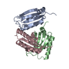
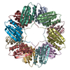
| Title | Populus tremula stable protein 1 with N-terminal binding peptide extension with hemin | ||||||
 Components Components | Stable protein 1 | ||||||
 Keywords Keywords | PLANT PROTEIN / Hemine / Protein engineering / Biohybrid / catalysis | ||||||
| Function / homology | pollen tube adhesion / Stress-response A/B barrel domain-containing protein HS1/DABB1-like / Stress responsive alpha-beta barrel / Stress responsive A/B Barrel Domain / Stress-response A/B barrel domain profile. / Stress responsive A/B Barrel Domain / Dimeric alpha-beta barrel / Stable protein 1 Function and homology information Function and homology information | ||||||
| Biological species |  | ||||||
| Method |  X-RAY DIFFRACTION / SYNCHROTRON / MOLECULAR REPLACEMENT / Resolution: 2.4 Å X-RAY DIFFRACTION / SYNCHROTRON / MOLECULAR REPLACEMENT / Resolution: 2.4 Å | ||||||
 Authors Authors | Sklyar, J. / Zeibaq, Y. / Bachar, O. / Yehezkeli, O. / Adir, N. | ||||||
| Funding support |  Israel, 1items Israel, 1items
| ||||||
 Citation Citation | Journal: Small Sci / Year: 2024 Title: A Bioengineered Stable Protein 1-Hemin Complex with Enhanced Peroxidase-Like Catalytic Properties. Authors: Zeibaq, Y. / Bachar, O. / Sklyar, J. / Adir, N. / Yehezkeli, O. | ||||||
| History |
|
- Structure visualization
| Structure viewer | Molecule: MolmilJmol/JSmol |
|---|
- Downloads & links
-Download
| PDBx/mmCIF format | 8ozs.cif.gz | 545.2 KB | Display | PDBx/mmCIF format |
|---|---|---|---|---|
| PDB format | pdb8ozs.ent.gz | 448.2 KB | Display | PDB format |
| PDBx/mmJSON format | 8ozs.json.gz | Tree view | PDBx/mmJSON format | |
| Others |  Other downloads Other downloads |
-Validation report
| Summary document | 8ozs_validation.pdf.gz | 607.3 KB | Display | wwPDB validaton report |
|---|---|---|---|---|
| Full document | 8ozs_full_validation.pdf.gz | 624.4 KB | Display | |
| Data in XML | 8ozs_validation.xml.gz | 104.6 KB | Display | |
| Data in CIF | 8ozs_validation.cif.gz | 148.7 KB | Display | |
| Arichive directory | https://data.pdbj.org/pub/pdb/validation_reports/oz/8ozsftp://data.pdbj.org/pub/pdb/validation_reports/oz/8ozs | HTTPS FTP |
-Related structure data
-Links
 PDBj
PDBj- Assembly
| Deposited unit | 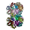
| ||||||||
|---|---|---|---|---|---|---|---|---|---|
| 1 | 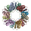
| ||||||||
| 2 | 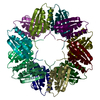
| ||||||||
| Unit cell |
|
-Components
| #1: Protein | Mass: 13681.366 Da / Num. of mol.: 24 Source method: isolated from a genetically manipulated source Source: (gene. exp.)  #2: Water | ChemComp-HOH / |  Mass: 18.015 Da / Num. of mol.: 1927 / Source method: isolated from a natural source / Formula: H2O Mass: 18.015 Da / Num. of mol.: 1927 / Source method: isolated from a natural source / Formula: H2OHas protein modification | N | |
|---|
-Experimental details
-Experiment
| Experiment | Method: X-RAY DIFFRACTION / Number of used crystals: 1 |
|---|
- Sample preparation
| Crystal | Density Matthews: 2.59 Å3/Da / Density % sol: 47.53 % |
|---|---|
| Crystal grow | Temperature: 293.15 K / Method: vapor diffusion, hanging drop Details: 7.5% v/v Tacsimate pH 7.0, 5% v/v 2-Propanol, 0.1M Imidazole pH 7.0, 13% w/v PEG 3350 |
-Data collection
| Diffraction | Mean temperature: 100 K / Serial crystal experiment: N |
|---|---|
| Diffraction source | Source: SYNCHROTRON / Site: ESRF  / Beamline: ID23-2 / Wavelength: 0.873128 Å / Beamline: ID23-2 / Wavelength: 0.873128 Å |
| Detector | Type: DECTRIS PILATUS3 2M / Detector: PIXEL / Date: Nov 26, 2022 |
| Radiation | Protocol: SINGLE WAVELENGTH / Monochromatic (M) / Laue (L): M / Scattering type: x-ray |
| Radiation wavelength | Wavelength: 0.873128 Å / Relative weight: 1 |
| Reflection | Resolution: 2.4→48.6 Å / Num. obs: 118649 / % possible obs: 99.8 % / Redundancy: 5.5 % / CC1/2: 0.996 / Rmerge(I) obs: 0.115 / Rrim(I) all: 0.14 / Net I/σ(I): 6.3 |
| Reflection shell | Resolution: 2.4→2.44 Å / Num. unique obs: 5863 / CC1/2: 0.955 |
- Processing
| Software |
| |||||||||||||||||||||||||||||||||||||||||||||||||||||||||||||||||||||||||||||||||||||||||||||||||||||||||||||||||||||||||||||||||||||||||||||||||||||||||||||||||||||||||||||||||||||||||||||||||||||||||||||||||||||||||
|---|---|---|---|---|---|---|---|---|---|---|---|---|---|---|---|---|---|---|---|---|---|---|---|---|---|---|---|---|---|---|---|---|---|---|---|---|---|---|---|---|---|---|---|---|---|---|---|---|---|---|---|---|---|---|---|---|---|---|---|---|---|---|---|---|---|---|---|---|---|---|---|---|---|---|---|---|---|---|---|---|---|---|---|---|---|---|---|---|---|---|---|---|---|---|---|---|---|---|---|---|---|---|---|---|---|---|---|---|---|---|---|---|---|---|---|---|---|---|---|---|---|---|---|---|---|---|---|---|---|---|---|---|---|---|---|---|---|---|---|---|---|---|---|---|---|---|---|---|---|---|---|---|---|---|---|---|---|---|---|---|---|---|---|---|---|---|---|---|---|---|---|---|---|---|---|---|---|---|---|---|---|---|---|---|---|---|---|---|---|---|---|---|---|---|---|---|---|---|---|---|---|---|---|---|---|---|---|---|---|---|---|---|---|---|---|---|---|---|
| Refinement | Method to determine structure: MOLECULAR REPLACEMENT / Resolution: 2.4→48.6 Å / SU ML: 0.28 / Cross valid method: FREE R-VALUE / σ(F): 1.34 / Phase error: 26.34 / Stereochemistry target values: ML
| |||||||||||||||||||||||||||||||||||||||||||||||||||||||||||||||||||||||||||||||||||||||||||||||||||||||||||||||||||||||||||||||||||||||||||||||||||||||||||||||||||||||||||||||||||||||||||||||||||||||||||||||||||||||||
| Solvent computation | Shrinkage radii: 0.9 Å / VDW probe radii: 1.1 Å / Solvent model: FLAT BULK SOLVENT MODEL | |||||||||||||||||||||||||||||||||||||||||||||||||||||||||||||||||||||||||||||||||||||||||||||||||||||||||||||||||||||||||||||||||||||||||||||||||||||||||||||||||||||||||||||||||||||||||||||||||||||||||||||||||||||||||
| Refinement step | Cycle: LAST / Resolution: 2.4→48.6 Å
| |||||||||||||||||||||||||||||||||||||||||||||||||||||||||||||||||||||||||||||||||||||||||||||||||||||||||||||||||||||||||||||||||||||||||||||||||||||||||||||||||||||||||||||||||||||||||||||||||||||||||||||||||||||||||
| Refine LS restraints |
| |||||||||||||||||||||||||||||||||||||||||||||||||||||||||||||||||||||||||||||||||||||||||||||||||||||||||||||||||||||||||||||||||||||||||||||||||||||||||||||||||||||||||||||||||||||||||||||||||||||||||||||||||||||||||
| LS refinement shell |
|
