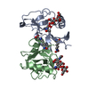
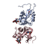
 Movie
Movie Controller


+ Open data

- Basic information
| Entry | Database: PDB / ID: 8ouq | |||||||||
|---|---|---|---|---|---|---|---|---|---|---|
| Title | Clr-11 from Rattus norvegicus | |||||||||
 Components Components | C-type lectin domain family 2 member D11 | |||||||||
 Keywords Keywords | IMMUNE SYSTEM / CTLD FOLD / NATURAL KILLER CELL | |||||||||
| Function / homology |  Function and homology information Function and homology informationnatural killer cell lectin-like receptor binding / carbohydrate binding / external side of plasma membrane Similarity search - Function | |||||||||
| Biological species |  | |||||||||
| Method |  X-RAY DIFFRACTION / SYNCHROTRON / MOLECULAR REPLACEMENT / Resolution: 1.6 Å X-RAY DIFFRACTION / SYNCHROTRON / MOLECULAR REPLACEMENT / Resolution: 1.6 Å | |||||||||
 Authors Authors | Skalova, T. / Blaha, J. / Kalouskova, B. / Skorepa, O. / Vanek, O. / Dohnalek, J. | |||||||||
| Funding support |  Czech Republic, European Union, 2items Czech Republic, European Union, 2items
| |||||||||
 Citation Citation | Journal: To Be Published Title: Clr-11 from Rattus norvegicus Authors: Skalova, T. / Blaha, J. / Kalouskova, B. / Skorepa, O. / Vanek, O. / Dohnalek, J. | |||||||||
| History |
|
- Structure visualization
| Structure viewer | Molecule: MolmilJmol/JSmol |
|---|
- Downloads & links
-Download
| PDBx/mmCIF format | 8ouq.cif.gz | 76.8 KB | Display | PDBx/mmCIF format |
|---|---|---|---|---|
| PDB format | pdb8ouq.ent.gz | Display | PDB format | |
| PDBx/mmJSON format | 8ouq.json.gz | Tree view | PDBx/mmJSON format | |
| Others |  Other downloads Other downloads |
-Validation report
| Arichive directory | https://data.pdbj.org/pub/pdb/validation_reports/ou/8ouqftp://data.pdbj.org/pub/pdb/validation_reports/ou/8ouq | HTTPS FTP |
|---|
-Related structure data
| Related structure data | |
|---|---|
| Similar structure data | |
| Experimental dataset #1 | Data reference: 10.15785/SBGRID/953 / Data set type: diffraction image data |
-Links
 PDBj
PDBj


- Assembly
| Deposited unit | 
| ||||||||
|---|---|---|---|---|---|---|---|---|---|
| 1 |
| ||||||||
| Unit cell |
|
-Components
| #1: Protein | Mass: 15729.235 Da / Num. of mol.: 2 Source method: isolated from a genetically manipulated source Source: (gene. exp.)  Homo sapiens (human) / Tissue (production host): HUMAN EMBRYONIC KIDNEY / References: UniProt: Q0H8B9 Homo sapiens (human) / Tissue (production host): HUMAN EMBRYONIC KIDNEY / References: UniProt: Q0H8B9#2: Sugar |   Type: D-saccharide, beta linking / Mass: 221.208 Da / Num. of mol.: 2 / Source method: obtained synthetically / Formula: C8H15NO6 / Feature type: SUBJECT OF INVESTIGATION Type: D-saccharide, beta linking / Mass: 221.208 Da / Num. of mol.: 2 / Source method: obtained synthetically / Formula: C8H15NO6 / Feature type: SUBJECT OF INVESTIGATION#3: Water | ChemComp-HOH / |  Mass: 18.015 Da / Num. of mol.: 276 / Source method: isolated from a natural source / Formula: H2O Mass: 18.015 Da / Num. of mol.: 276 / Source method: isolated from a natural source / Formula: H2OHas ligand of interest | Y | Has protein modification | Y | |
|---|
-Experimental details
-Experiment
| Experiment | Method: X-RAY DIFFRACTION / Number of used crystals: 1 |
|---|
- Sample preparation
| Crystal | Density Matthews: 2.11 Å3/Da / Density % sol: 42 % |
|---|---|
| Crystal grow | Temperature: 293 K / Method: vapor diffusion, sitting drop / pH: 5 / Details: 2 M sodium formate, 0.1 M sodium acetate, pH 5.0 |
-Data collection
| Diffraction | Mean temperature: 100 K / Serial crystal experiment: N |
|---|---|
| Diffraction source | Source: SYNCHROTRON / Site: Diamond  / Beamline: I04-1 / Wavelength: 0.97949 Å / Beamline: I04-1 / Wavelength: 0.97949 Å |
| Detector | Type: DECTRIS PILATUS 6M / Detector: PIXEL / Date: Feb 3, 2016 |
| Radiation | Protocol: SINGLE WAVELENGTH / Monochromatic (M) / Laue (L): M / Scattering type: x-ray |
| Radiation wavelength | Wavelength: 0.97949 Å / Relative weight: 1 |
| Reflection | Resolution: 1.6→46.23 Å / Num. obs: 34197 / % possible obs: 99.1 % / Redundancy: 13.8 % / Biso Wilson estimate: 16.1 Å2 / CC1/2: 0.995 / Rmerge(I) obs: 0.116 / Rpim(I) all: 0.032 / Rrim(I) all: 0.12 / Χ2: 0.98 / Net I/σ(I): 11 |
| Reflection shell | Resolution: 1.6→1.63 Å / Redundancy: 13.9 % / Rmerge(I) obs: 0.933 / Mean I/σ(I) obs: 2.7 / Num. unique obs: 1687 / CC1/2: 0.967 / Rpim(I) all: 0.258 / Rrim(I) all: 0.968 / Χ2: 0.99 / % possible all: 97.1 |
- Processing
| Software |
| |||||||||||||||||||||||||||||||||||||||||||||||||||||||||||||||||||||||||||||||||||||||||||||||||||||||||||||||||||||||||||||||||||||||||||||||||||||||||||
|---|---|---|---|---|---|---|---|---|---|---|---|---|---|---|---|---|---|---|---|---|---|---|---|---|---|---|---|---|---|---|---|---|---|---|---|---|---|---|---|---|---|---|---|---|---|---|---|---|---|---|---|---|---|---|---|---|---|---|---|---|---|---|---|---|---|---|---|---|---|---|---|---|---|---|---|---|---|---|---|---|---|---|---|---|---|---|---|---|---|---|---|---|---|---|---|---|---|---|---|---|---|---|---|---|---|---|---|---|---|---|---|---|---|---|---|---|---|---|---|---|---|---|---|---|---|---|---|---|---|---|---|---|---|---|---|---|---|---|---|---|---|---|---|---|---|---|---|---|---|---|---|---|---|---|---|---|
| Refinement | Method to determine structure: MOLECULAR REPLACEMENT / Resolution: 1.6→46.23 Å / Cor.coef. Fo:Fc: 0.967 / SU B: 1.993 / SU ML: 0.067 / Cross valid method: THROUGHOUT / ESU R: 0.096 Details: Hydrogens have been added in their riding positions
| |||||||||||||||||||||||||||||||||||||||||||||||||||||||||||||||||||||||||||||||||||||||||||||||||||||||||||||||||||||||||||||||||||||||||||||||||||||||||||
| Solvent computation | Ion probe radii: 0.8 Å / Shrinkage radii: 0.8 Å / VDW probe radii: 1.2 Å / Solvent model: MASK BULK SOLVENT | |||||||||||||||||||||||||||||||||||||||||||||||||||||||||||||||||||||||||||||||||||||||||||||||||||||||||||||||||||||||||||||||||||||||||||||||||||||||||||
| Displacement parameters | Biso mean: 20.127 Å2
| |||||||||||||||||||||||||||||||||||||||||||||||||||||||||||||||||||||||||||||||||||||||||||||||||||||||||||||||||||||||||||||||||||||||||||||||||||||||||||
| Refinement step | Cycle: LAST / Resolution: 1.6→46.23 Å
| |||||||||||||||||||||||||||||||||||||||||||||||||||||||||||||||||||||||||||||||||||||||||||||||||||||||||||||||||||||||||||||||||||||||||||||||||||||||||||
| Refine LS restraints |
| |||||||||||||||||||||||||||||||||||||||||||||||||||||||||||||||||||||||||||||||||||||||||||||||||||||||||||||||||||||||||||||||||||||||||||||||||||||||||||
| LS refinement shell |
|
