 Movie
Movie Controller

[English] 日本語
 Yorodumi
- PDB-8ota: High resolution crystal structure of a Leaf-branch compost cutina... -
+ Open data

- Basic information
| Entry | Database: PDB / ID: 8ota | ||||||
|---|---|---|---|---|---|---|---|
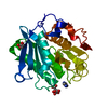
| Title | High resolution crystal structure of a Leaf-branch compost cutinase quadruple variant | ||||||
 Components Components | Leaf-branch compost cutinase | ||||||
 Keywords Keywords | HYDROLASE / SERINE ESTERASE / CUTINASE | ||||||
| Function / homology |  Function and homology information Function and homology informationacetylesterase activity / poly(ethylene terephthalate) hydrolase / cutinase / cutinase activity / extracellular region Similarity search - Function | ||||||
| Biological species |  uncultured bacterium (environmental samples) uncultured bacterium (environmental samples) | ||||||
| Method |  X-RAY DIFFRACTION / SYNCHROTRON / MOLECULAR REPLACEMENT / Resolution: 1.72 Å X-RAY DIFFRACTION / SYNCHROTRON / MOLECULAR REPLACEMENT / Resolution: 1.72 Å | ||||||
 Authors Authors | Gavalda, S. / Cioci, G. / Marty, A. / Duquesne, S. | ||||||
| Funding support |  France, 1items France, 1items
| ||||||
 Citation Citation | Journal: Biophys.J. / Year: 2024 Title: Exploring the pH dependence of an improved PETase. Authors: Charlier, C. / Gavalda, S. / Grga, J. / Perrot, L. / Gabrielli, V. / Lohr, F. / Schorghuber, J. / Lichtenecker, R. / Arnal, G. / Marty, A. / Tournier, V. / Lippens, G. | ||||||
| History |
|
- Structure visualization
| Structure viewer | Molecule: MolmilJmol/JSmol |
|---|
- Downloads & links
-Download
| PDBx/mmCIF format | 8ota.cif.gz | 88 KB | Display | PDBx/mmCIF format |
|---|---|---|---|---|
| PDB format | pdb8ota.ent.gz | 51.6 KB | Display | PDB format |
| PDBx/mmJSON format | 8ota.json.gz | Tree view | PDBx/mmJSON format | |
| Others |  Other downloads Other downloads |
-Validation report
| Arichive directory | https://data.pdbj.org/pub/pdb/validation_reports/ot/8otaftp://data.pdbj.org/pub/pdb/validation_reports/ot/8ota | HTTPS FTP |
|---|
-Related structure data
| Related structure data | |
|---|---|
| Similar structure data |
-Links
 PDBj
PDBj
- Assembly
| Deposited unit | 
| ||||||||||||
|---|---|---|---|---|---|---|---|---|---|---|---|---|---|
| 1 |
| ||||||||||||
| Unit cell |
| ||||||||||||
| Components on special symmetry positions |
|
-Components
| #1: Protein | Mass: 27795.168 Da / Num. of mol.: 1 / Mutation: F243I, D238C, S283C, Y127G Source method: isolated from a genetically manipulated source Source: (gene. exp.) uncultured bacterium (environmental samples)Production host: References: UniProt: G9BY57, cutinase, poly(ethylene terephthalate) hydrolase |
|---|---|
| #2: Chemical | ChemComp-DIO /   Mass: 88.105 Da / Num. of mol.: 1 / Source method: obtained synthetically / Formula: C4H8O2 Mass: 88.105 Da / Num. of mol.: 1 / Source method: obtained synthetically / Formula: C4H8O2 |
| #3: Water | ChemComp-HOH /  Mass: 18.015 Da / Num. of mol.: 369 / Source method: isolated from a natural source / Formula: H2O Mass: 18.015 Da / Num. of mol.: 369 / Source method: isolated from a natural source / Formula: H2O |
| Has ligand of interest | N |
| Has protein modification | Y |
-Experimental details
-Experiment
| Experiment | Method: X-RAY DIFFRACTION / Number of used crystals: 1 |
|---|
- Sample preparation
| Crystal | Density Matthews: 2.24 Å3/Da / Density % sol: 45.17 % |
|---|---|
| Crystal grow | Temperature: 285 K / Method: vapor diffusion, sitting drop / Details: 0.1 M Bicine pH9, 10% PEG20000, 2% dioxane |
-Data collection
| Diffraction | Mean temperature: 100 K / Serial crystal experiment: N |
|---|---|
| Diffraction source | Source: SYNCHROTRON / Site: ESRF / Beamline: MASSIF-3 / Wavelength: 0.9677 Å |
| Detector | Type: DECTRIS EIGER X 4M / Detector: PIXEL / Date: Oct 12, 2020 |
| Radiation | Protocol: SINGLE WAVELENGTH / Monochromatic (M) / Laue (L): M / Scattering type: x-ray |
| Radiation wavelength | Wavelength: 0.9677 Å / Relative weight: 1 |
| Reflection | Resolution: 1.719→36.07 Å / Num. obs: 26667 / % possible obs: 100 % / Redundancy: 9.8 % / Biso Wilson estimate: 19.53 Å2 / CC1/2: 0.997 / Net I/σ(I): 12.9 |
| Reflection shell | Resolution: 1.719→1.749 Å / Redundancy: 6.4 % / Rmerge(I) obs: 0.63 / Mean I/σ(I) obs: 2.3 / Num. unique obs: 1350 / CC1/2: 0.774 / Rpim(I) all: 0.273 / Rrim(I) all: 0.706 / % possible all: 100 |
- Processing
| Software |
| |||||||||||||||||||||||||||||||||||||||||||||||||||||||||||||||||||||||||||||
|---|---|---|---|---|---|---|---|---|---|---|---|---|---|---|---|---|---|---|---|---|---|---|---|---|---|---|---|---|---|---|---|---|---|---|---|---|---|---|---|---|---|---|---|---|---|---|---|---|---|---|---|---|---|---|---|---|---|---|---|---|---|---|---|---|---|---|---|---|---|---|---|---|---|---|---|---|---|---|
| Refinement | Method to determine structure: MOLECULAR REPLACEMENT / Resolution: 1.72→36.07 Å / SU ML: 0.2448 / Cross valid method: FREE R-VALUE / σ(F): 1.39 / Phase error: 19.4438 Stereochemistry target values: GeoStd + Monomer Library + CDL v1.2
| |||||||||||||||||||||||||||||||||||||||||||||||||||||||||||||||||||||||||||||
| Solvent computation | Shrinkage radii: 0.9 Å / VDW probe radii: 1.11 Å / Solvent model: FLAT BULK SOLVENT MODEL | |||||||||||||||||||||||||||||||||||||||||||||||||||||||||||||||||||||||||||||
| Displacement parameters | Biso mean: 18.22 Å2 | |||||||||||||||||||||||||||||||||||||||||||||||||||||||||||||||||||||||||||||
| Refinement step | Cycle: LAST / Resolution: 1.72→36.07 Å
| |||||||||||||||||||||||||||||||||||||||||||||||||||||||||||||||||||||||||||||
| Refine LS restraints |
| |||||||||||||||||||||||||||||||||||||||||||||||||||||||||||||||||||||||||||||
| LS refinement shell |
|
