 Movie
Movie Controller

[English] 日本語
 Yorodumi
- PDB-8orp: Crystal structure of Drosophila melanogaster alpha-amylase in com... -
+ Open data

- Basic information
| Entry | Database: PDB / ID: 8orp | ||||||
|---|---|---|---|---|---|---|---|
| Title | Crystal structure of Drosophila melanogaster alpha-amylase in complex with the inhibitor acarbose | ||||||
 Components Components | Alpha-amylase A | ||||||
 Keywords Keywords | HYDROLASE / ALPHA-AMYLASE / ALPHA-1 / 4-GLUCAN-4-GLUCANOHYDROLASE / BETA- ALPHA-EIGHT BARREL / ADAPTATION / INHIBITOR / ACARBOSE | ||||||
| Function / homology |  Function and homology information Function and homology informationDigestion of dietary carbohydrate / alpha-amylase / alpha-amylase activity / carbohydrate catabolic process / carbohydrate metabolic process / calcium ion binding / extracellular space / extracellular region Similarity search - Function | ||||||
| Biological species |  | ||||||
| Method |  X-RAY DIFFRACTION / SYNCHROTRON / FOURIER SYNTHESIS / Resolution: 2 Å X-RAY DIFFRACTION / SYNCHROTRON / FOURIER SYNTHESIS / Resolution: 2 Å | ||||||
 Authors Authors | Aghajari, N. / Haser, R. | ||||||
| Funding support |  France, 1items France, 1items
| ||||||
 Citation Citation | Journal: Molecules / Year: 2023 Title: Structural and Functional Characterization of Drosophila melanogaster alpha-Amylase. Authors: Rhimi, M. / Da Lage, J.L. / Haser, R. / Feller, G. / Aghajari, N. | ||||||
| History |
|
- Structure visualization
| Structure viewer | Molecule: MolmilJmol/JSmol |
|---|
- Downloads & links
-Download
| PDBx/mmCIF format | 8orp.cif.gz | 275.8 KB | Display | PDBx/mmCIF format |
|---|---|---|---|---|
| PDB format | pdb8orp.ent.gz | 176.6 KB | Display | PDB format |
| PDBx/mmJSON format | 8orp.json.gz | Tree view | PDBx/mmJSON format | |
| Others |  Other downloads Other downloads |
-Validation report
| Summary document | 8orp_validation.pdf.gz | 4.2 MB | Display | wwPDB validaton report |
|---|---|---|---|---|
| Full document | 8orp_full_validation.pdf.gz | 4.1 MB | Display | |
| Data in XML | 8orp_validation.xml.gz | 51.5 KB | Display | |
| Data in CIF | 8orp_validation.cif.gz | 73.3 KB | Display | |
| Arichive directory | https://data.pdbj.org/pub/pdb/validation_reports/or/8orpftp://data.pdbj.org/pub/pdb/validation_reports/or/8orp | HTTPS FTP |
-Related structure data

-Links
 PDBj
PDBj


- Assembly
| Deposited unit | 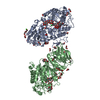
| |||||||||||||||||||||||||||||||||||||||||||||||||||||||||||||||||||||||||||||||
|---|---|---|---|---|---|---|---|---|---|---|---|---|---|---|---|---|---|---|---|---|---|---|---|---|---|---|---|---|---|---|---|---|---|---|---|---|---|---|---|---|---|---|---|---|---|---|---|---|---|---|---|---|---|---|---|---|---|---|---|---|---|---|---|---|---|---|---|---|---|---|---|---|---|---|---|---|---|---|---|---|
| 1 | 
| |||||||||||||||||||||||||||||||||||||||||||||||||||||||||||||||||||||||||||||||
| 2 | 
| |||||||||||||||||||||||||||||||||||||||||||||||||||||||||||||||||||||||||||||||
| Unit cell |
| |||||||||||||||||||||||||||||||||||||||||||||||||||||||||||||||||||||||||||||||
| Noncrystallographic symmetry (NCS) | NCS domain:
NCS domain segments: Ens-ID: ens_1
|
-Components
-Protein , 1 types, 2 molecules AB
| #1: Protein | Mass: 53797.332 Da / Num. of mol.: 2 Source method: isolated from a genetically manipulated source Source: (gene. exp.)  |
|---|
-Sugars , 2 types, 4 molecules
| #2: Polysaccharide | Type: oligosaccharide / Mass: 647.622 Da / Num. of mol.: 2 / Source method: obtained synthetically #3: Polysaccharide | Type: oligosaccharide / Mass: 485.481 Da / Num. of mol.: 2 / Source method: obtained synthetically |
|---|
-Non-polymers , 4 types, 919 molecules 


| #4: Chemical |  Mass: 35.453 Da / Num. of mol.: 2 / Source method: obtained synthetically / Formula: Cl / Feature type: SUBJECT OF INVESTIGATION Mass: 35.453 Da / Num. of mol.: 2 / Source method: obtained synthetically / Formula: Cl / Feature type: SUBJECT OF INVESTIGATION#5: Chemical | ChemComp-EDO /  Mass: 62.068 Da / Num. of mol.: 38 / Source method: obtained synthetically / Formula: C2H6O2 Mass: 62.068 Da / Num. of mol.: 38 / Source method: obtained synthetically / Formula: C2H6O2#6: Chemical | ChemComp-SR /  Mass: 87.620 Da / Num. of mol.: 4 / Source method: obtained synthetically / Formula: Sr / Feature type: SUBJECT OF INVESTIGATION Mass: 87.620 Da / Num. of mol.: 4 / Source method: obtained synthetically / Formula: Sr / Feature type: SUBJECT OF INVESTIGATION#7: Water | ChemComp-HOH / | Mass: 18.015 Da / Num. of mol.: 875 / Source method: isolated from a natural source / Formula: H2O |
|---|
-Details
| Has ligand of interest | Y |
|---|---|
| Has protein modification | Y |
-Experimental details
-Experiment
| Experiment | Method: X-RAY DIFFRACTION / Number of used crystals: 1 |
|---|
- Sample preparation
| Crystal | Density Matthews: 2.62 Å3/Da / Density % sol: 53.1 % |
|---|---|
| Crystal grow | Temperature: 291 K / Method: vapor diffusion, hanging drop Details: 20 % (v/v) PEG monomethyl ether 550, 0.1 M sodium chloride, 0.1 M bicine buffer (pH 9.0) and 10 mM strontium chloride. Soak in a solution containing 10 mM acarbose. |
-Data collection
| Diffraction | Mean temperature: 100 K / Serial crystal experiment: N |
|---|---|
| Diffraction source | Source: SYNCHROTRON / Site: ESRF / Beamline: ID14-1 / Wavelength: 0.9334 Å |
| Detector | Type: ADSC QUANTUM 210 / Detector: CCD / Date: Feb 7, 2009 |
| Radiation | Monochromator: diamond 111 / Protocol: SINGLE WAVELENGTH / Monochromatic (M) / Laue (L): M / Scattering type: x-ray |
| Radiation wavelength | Wavelength: 0.9334 Å / Relative weight: 1 |
| Reflection | Resolution: 2→29.6 Å / Num. obs: 75240 / % possible obs: 99.8 % / Redundancy: 3.7 % / Biso Wilson estimate: 17.57 Å2 / CC1/2: 0.993 / Net I/σ(I): 9 |
| Reflection shell | Resolution: 2→2.1 Å / Num. unique obs: 10118 / CC1/2: 0.778 / % possible all: 99.7 |
- Processing
| Software |
| ||||||||||||||||||||||||||||||||||||||||||||||||||||||||||||||||||||||||||||||||||||||||||||||||||||||||||||||||||||||||||||||||||||||||||||||||||||||||||||||||||||||||||||||||||||||||||||||||||||
|---|---|---|---|---|---|---|---|---|---|---|---|---|---|---|---|---|---|---|---|---|---|---|---|---|---|---|---|---|---|---|---|---|---|---|---|---|---|---|---|---|---|---|---|---|---|---|---|---|---|---|---|---|---|---|---|---|---|---|---|---|---|---|---|---|---|---|---|---|---|---|---|---|---|---|---|---|---|---|---|---|---|---|---|---|---|---|---|---|---|---|---|---|---|---|---|---|---|---|---|---|---|---|---|---|---|---|---|---|---|---|---|---|---|---|---|---|---|---|---|---|---|---|---|---|---|---|---|---|---|---|---|---|---|---|---|---|---|---|---|---|---|---|---|---|---|---|---|---|---|---|---|---|---|---|---|---|---|---|---|---|---|---|---|---|---|---|---|---|---|---|---|---|---|---|---|---|---|---|---|---|---|---|---|---|---|---|---|---|---|---|---|---|---|---|---|---|---|
| Refinement | Method to determine structure: FOURIER SYNTHESIS / Resolution: 2→29.57 Å / SU ML: 0.2105 / Cross valid method: FREE R-VALUE / σ(F): 1.36 / Phase error: 20.9939 Stereochemistry target values: GeoStd + Monomer Library + CDL v1.2
| ||||||||||||||||||||||||||||||||||||||||||||||||||||||||||||||||||||||||||||||||||||||||||||||||||||||||||||||||||||||||||||||||||||||||||||||||||||||||||||||||||||||||||||||||||||||||||||||||||||
| Solvent computation | Shrinkage radii: 0.9 Å / VDW probe radii: 1.1 Å / Solvent model: FLAT BULK SOLVENT MODEL | ||||||||||||||||||||||||||||||||||||||||||||||||||||||||||||||||||||||||||||||||||||||||||||||||||||||||||||||||||||||||||||||||||||||||||||||||||||||||||||||||||||||||||||||||||||||||||||||||||||
| Displacement parameters | Biso mean: 18.94 Å2 | ||||||||||||||||||||||||||||||||||||||||||||||||||||||||||||||||||||||||||||||||||||||||||||||||||||||||||||||||||||||||||||||||||||||||||||||||||||||||||||||||||||||||||||||||||||||||||||||||||||
| Refinement step | Cycle: LAST / Resolution: 2→29.57 Å
| ||||||||||||||||||||||||||||||||||||||||||||||||||||||||||||||||||||||||||||||||||||||||||||||||||||||||||||||||||||||||||||||||||||||||||||||||||||||||||||||||||||||||||||||||||||||||||||||||||||
| Refine LS restraints |
| ||||||||||||||||||||||||||||||||||||||||||||||||||||||||||||||||||||||||||||||||||||||||||||||||||||||||||||||||||||||||||||||||||||||||||||||||||||||||||||||||||||||||||||||||||||||||||||||||||||
| Refine LS restraints NCS | Type: Torsion NCS / Rms dev position: 0.461429737109 Å | ||||||||||||||||||||||||||||||||||||||||||||||||||||||||||||||||||||||||||||||||||||||||||||||||||||||||||||||||||||||||||||||||||||||||||||||||||||||||||||||||||||||||||||||||||||||||||||||||||||
| LS refinement shell |
|
