Movie
Movie Controller

[English] 日本語
 Yorodumi
- PDB-8orh: Knockout of GMC-oxidoreductase genes reveals that functional redu... -
+ Open data

- Basic information
| Entry | Database: PDB / ID: 8orh | ||||||
|---|---|---|---|---|---|---|---|
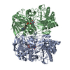
| Title | Knockout of GMC-oxidoreductase genes reveals that functional redundancy preserves mimivirus essential functions | ||||||
 Components Components | Putative GMC-type oxidoreductase | ||||||
 Keywords Keywords | STRUCTURAL PROTEIN / GMC-oxidoreductase / genomic fiber / mimivirus | ||||||
| Function / homology |  Function and homology information Function and homology informationoxidoreductase activity, acting on CH-OH group of donors / flavin adenine dinucleotide binding Similarity search - Function | ||||||
| Biological species |  Mimivirus reunion Mimivirus reunion | ||||||
| Method | ELECTRON MICROSCOPY / helical reconstruction / cryo EM / Resolution: 4.2 Å | ||||||
 Authors Authors | Alempic, J.M. / Bisio, H. / Villalta, A. / Santini, S. / Lartigue, A. / Schmitt, A. / Bugnot, C. / Notaro, A. / Belmudes, L. / Adrait, A. ...Alempic, J.M. / Bisio, H. / Villalta, A. / Santini, S. / Lartigue, A. / Schmitt, A. / Bugnot, C. / Notaro, A. / Belmudes, L. / Adrait, A. / Poirot, O. / Ptchelkine, D. / De Castro, C. / Coute, Y. / Abergel, C. | ||||||
| Funding support | European Union, 1items
| ||||||
 Citation Citation | Journal: Microlife / Year: 2024 Title: Functional redundancy revealed by the deletion of the mimivirus GMC-oxidoreductase genes. Authors: Jean-Marie Alempic / Hugo Bisio / Alejandro Villalta / Sébastien Santini / Audrey Lartigue / Alain Schmitt / Claire Bugnot / Anna Notaro / Lucid Belmudes / Annie Adrait / Olivier Poirot / ...Authors: Jean-Marie Alempic / Hugo Bisio / Alejandro Villalta / Sébastien Santini / Audrey Lartigue / Alain Schmitt / Claire Bugnot / Anna Notaro / Lucid Belmudes / Annie Adrait / Olivier Poirot / Denis Ptchelkine / Cristina De Castro / Yohann Couté / Chantal Abergel /   Abstract: The mimivirus 1.2 Mb genome was shown to be organized into a nucleocapsid-like genomic fiber encased in the nucleoid compartment inside the icosahedral capsid. The genomic fiber protein shell is ...The mimivirus 1.2 Mb genome was shown to be organized into a nucleocapsid-like genomic fiber encased in the nucleoid compartment inside the icosahedral capsid. The genomic fiber protein shell is composed of a mixture of two GMC-oxidoreductase paralogs, one of them being the main component of the glycosylated layer of fibrils at the surface of the virion. In this study, we determined the effect of the deletion of each of the corresponding genes on the genomic fiber and the layer of surface fibrils. First, we deleted the GMC-oxidoreductase, the most abundant in the genomic fiber, and determined its structure and composition in the mutant. As expected, it was composed of the second GMC-oxidoreductase and contained 5- and 6-start helices similar to the wild-type fiber. This result led us to propose a model explaining their coexistence. Then we deleted the GMC-oxidoreductase, the most abundant in the layer of fibrils, to analyze its protein composition in the mutant. Second, we showed that the fitness of single mutants and the double mutant were not decreased compared with the wild-type viruses under laboratory conditions. Third, we determined that deleting the GMC-oxidoreductase genes did not impact the glycosylation or the glycan composition of the layer of surface fibrils, despite modifying their protein composition. Because the glycosylation machinery and glycan composition of members of different clades are different, we expanded the analysis of the protein composition of the layer of fibrils to members of the B and C clades and showed that it was different among the three clades and even among isolates within the same clade. Taken together, the results obtained on two distinct central processes (genome packaging and virion coating) illustrate an unexpected functional redundancy in members of the family , suggesting this may be the major evolutionary force behind their giant genomes. | ||||||
| History |
|
- Structure visualization
| Structure viewer | Molecule: MolmilJmol/JSmol |
|---|
- Downloads & links
-Download
| PDBx/mmCIF format | 8orh.cif.gz | 230.5 KB | Display | PDBx/mmCIF format |
|---|---|---|---|---|
| PDB format | pdb8orh.ent.gz | 185.4 KB | Display | PDB format |
| PDBx/mmJSON format | 8orh.json.gz | Tree view | PDBx/mmJSON format | |
| Others |  Other downloads Other downloads |
-Validation report
| Arichive directory | https://data.pdbj.org/pub/pdb/validation_reports/or/8orhftp://data.pdbj.org/pub/pdb/validation_reports/or/8orh | HTTPS FTP |
|---|
-Related structure data
| Related structure data |  17125MC  8orsC M: map data used to model this data C: citing same article ( |
|---|---|
| Similar structure data |

-Links
 PDBj
PDBj
- Assembly
| Deposited unit | 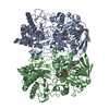
|
|---|---|
| 1 |
|
-Components
| #1: Protein | Mass: 77018.023 Da / Num. of mol.: 2 / Source method: isolated from a natural source Details: soruce organism: mimivirus reunion mutant where the qu_946 gene has been replaced by a selection cassette. Source: (natural) Mimivirus reunion / Variant: KO_qu946 / Strain: HB1931 / References: UniProt: A0A8A5IZP6#2: Chemical |   Mass: 785.550 Da / Num. of mol.: 2 / Source method: obtained synthetically / Formula: C27H33N9O15P2 / Feature type: SUBJECT OF INVESTIGATION / Comment: FAD*YM Mass: 785.550 Da / Num. of mol.: 2 / Source method: obtained synthetically / Formula: C27H33N9O15P2 / Feature type: SUBJECT OF INVESTIGATION / Comment: FAD*YMHas ligand of interest | Y | |
|---|
-Experimental details
-Experiment
| Experiment | Method: ELECTRON MICROSCOPY |
|---|---|
| EM experiment | Aggregation state: FILAMENT / 3D reconstruction method: helical reconstruction |
- Sample preparation
| Component | Name: Mimivirus reunion / Type: VIRUS Details: in the source organism, qu_946 gene has been removed and replaced by a selection cassette. Entity ID: #1 / Source: NATURAL |
|---|---|
| Molecular weight | Experimental value: NO |
| Source (natural) | Organism: Mimivirus reunion |
| Details of virus | Empty: NO / Enveloped: NO / Isolate: STRAIN / Type: VIRUS-LIKE PARTICLE |
| Natural host | Organism: Acanthamoeba castellanii str. Neff / Strain: KO_qu946 |
| Virus shell | Name: genomic fiber / Diameter: 320 nm |
| Buffer solution | pH: 7.5 |
| Buffer component | Conc.: 40 mmol/l / Name: Tris-HCl / Formula: Tris-HCl |
| Specimen | Embedding applied: NO / Shadowing applied: NO / Staining applied: NO / Vitrification applied: YES / Details: genomic fiber purified on ClCs gradient |
| Specimen support | Grid material: COPPER / Grid type: Quantifoil R2/1 |
| Vitrification | Instrument: FEI VITROBOT MARK IV / Cryogen name: ETHANE-PROPANE / Humidity: 100 % / Chamber temperature: 277 K |
- Electron microscopy imaging
| Experimental equipment |  Model: Titan Krios / Image courtesy: FEI Company |
|---|---|
| Microscopy | Model: FEI TITAN KRIOS |
| Electron gun | Electron source:  FIELD EMISSION GUN / Accelerating voltage: 300 kV / Illumination mode: OTHER FIELD EMISSION GUN / Accelerating voltage: 300 kV / Illumination mode: OTHER |
| Electron lens | Mode: BRIGHT FIELD / Nominal magnification: 81000 X / Nominal defocus max: 2800 nm / Nominal defocus min: 600 nm / Cs: 2.7 mm / C2 aperture diameter: 50 µm |
| Specimen holder | Cryogen: NITROGEN |
| Image recording | Average exposure time: 2.3 sec. / Electron dose: 35.597 e/Å2 / Film or detector model: GATAN K3 (6k x 4k) / Num. of real images: 2224 |
| EM imaging optics | Energyfilter name: GIF Bioquantum |
| Image scans | Width: 5760 / Height: 4092 |
- Processing
| Software | Name: PHENIX / Version: 1.20.1_4487: / Classification: refinement | ||||||||||||||||||||||||||||||||||||||||
|---|---|---|---|---|---|---|---|---|---|---|---|---|---|---|---|---|---|---|---|---|---|---|---|---|---|---|---|---|---|---|---|---|---|---|---|---|---|---|---|---|---|
| EM software |
| ||||||||||||||||||||||||||||||||||||||||
| CTF correction | Type: PHASE FLIPPING AND AMPLITUDE CORRECTION | ||||||||||||||||||||||||||||||||||||||||
| Helical symmerty | Angular rotation/subunit: 49.426 ° / Axial rise/subunit: 20.497 Å / Axial symmetry: C3 | ||||||||||||||||||||||||||||||||||||||||
| Particle selection | Num. of particles selected: 172431 | ||||||||||||||||||||||||||||||||||||||||
| 3D reconstruction | Resolution: 4.2 Å / Resolution method: FSC 0.5 CUT-OFF / Num. of particles: 20899 / Num. of class averages: 2 / Symmetry type: HELICAL | ||||||||||||||||||||||||||||||||||||||||
| Atomic model building | B value: 40.56 / Protocol: RIGID BODY FIT / Space: REAL | ||||||||||||||||||||||||||||||||||||||||
| Atomic model building | PDB-ID: 4Z24 Accession code: 4Z24 / Chain residue range: 4-652 / Pdb chain residue range: 4-652 / Source name: PDB / Type: experimental model | ||||||||||||||||||||||||||||||||||||||||
| Refine LS restraints |
|