 Movie
Movie Controller

[English] 日本語
 Yorodumi
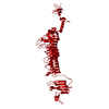
- PDB-8oq1: Crystal structure of tailspike depolymerase (APK14_gp49) from Aci... -
+ Open data

- Basic information
| Entry | Database: PDB / ID: 8oq1 | ||||||
|---|---|---|---|---|---|---|---|
| Title | Crystal structure of tailspike depolymerase (APK14_gp49) from Acinetobacter phage vB_AbaP_APK14 | ||||||
 Components Components | Tail spike protein, structural depolymerase | ||||||
 Keywords Keywords | VIRAL PROTEIN / bacteriophage / Acinetobacter baumannii / tailspike depolymerase / Acinetobacter phage | ||||||
| Function / homology | symbiont entry into host cell via disruption of host cell glycocalyx / symbiont entry into host cell via disruption of host cell envelope / virus tail / Pectin lyase fold / Pectin lyase fold/virulence factor / DI(HYDROXYETHYL)ETHER / Tail spike protein, structural depolymerase Function and homology information Function and homology information | ||||||
| Biological species |  Acinetobacter phage vB_AbaP_APK14 (virus) Acinetobacter phage vB_AbaP_APK14 (virus) | ||||||
| Method |  X-RAY DIFFRACTION / SYNCHROTRON / SAD / Resolution: 1.55 Å X-RAY DIFFRACTION / SYNCHROTRON / SAD / Resolution: 1.55 Å | ||||||
 Authors Authors | Matyuta, I.O. / Boyko, K.M. / Nikolaeva, A.Y. / Shneider, M.M. / Timoshina, O.Y. / Miroshnikov, K.A. / Popov, V.O. | ||||||
| Funding support |  Russian Federation, 1items Russian Federation, 1items
| ||||||
 Citation Citation | Journal: Int J Mol Sci / Year: 2023 Title: Friunavirus Phage-Encoded Depolymerases Specific to Different Capsular Types of Acinetobacter baumannii. Authors: Timoshina, O.Y. / Kasimova, A.A. / Shneider, M.M. / Matyuta, I.O. / Nikolaeva, A.Y. / Evseev, P.V. / Arbatsky, N.P. / Shashkov, A.S. / Chizhov, A.O. / Shelenkov, A.A. / Mikhaylova, Y.V. / ...Authors: Timoshina, O.Y. / Kasimova, A.A. / Shneider, M.M. / Matyuta, I.O. / Nikolaeva, A.Y. / Evseev, P.V. / Arbatsky, N.P. / Shashkov, A.S. / Chizhov, A.O. / Shelenkov, A.A. / Mikhaylova, Y.V. / Slukin, P.V. / Volozhantsev, N.V. / Boyko, K.M. / Knirel, Y.A. / Miroshnikov, K.A. / Popova, A.V. | ||||||
| History |
|
- Structure visualization
| Structure viewer | Molecule: MolmilJmol/JSmol |
|---|
- Downloads & links
-Download
| PDBx/mmCIF format | 8oq1.cif.gz | 161.7 KB | Display | PDBx/mmCIF format |
|---|---|---|---|---|
| PDB format | pdb8oq1.ent.gz | Display | PDB format | |
| PDBx/mmJSON format | 8oq1.json.gz | Tree view | PDBx/mmJSON format | |
| Others |  Other downloads Other downloads |
-Validation report
| Arichive directory | https://data.pdbj.org/pub/pdb/validation_reports/oq/8oq1ftp://data.pdbj.org/pub/pdb/validation_reports/oq/8oq1 | HTTPS FTP |
|---|
-Related structure data

-Links
 PDBj
PDBj- Assembly
| Deposited unit | 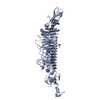
| ||||||||||||||||||||||||||||||
|---|---|---|---|---|---|---|---|---|---|---|---|---|---|---|---|---|---|---|---|---|---|---|---|---|---|---|---|---|---|---|---|
| 1 | 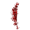
| ||||||||||||||||||||||||||||||
| Unit cell |
| ||||||||||||||||||||||||||||||
| Components on special symmetry positions |
|
-Components
| #1: Protein | Mass: 79618.969 Da / Num. of mol.: 1 Source method: isolated from a genetically manipulated source Source: (gene. exp.) Acinetobacter phage vB_AbaP_APK14 (virus)Gene: ABPK482_gp49 / Production host:  |
|---|---|
| #2: Chemical | ChemComp-PEG /   Mass: 106.120 Da / Num. of mol.: 1 / Source method: obtained synthetically / Formula: C4H10O3 Mass: 106.120 Da / Num. of mol.: 1 / Source method: obtained synthetically / Formula: C4H10O3 |
| #3: Chemical | ChemComp-GOL /   Mass: 92.094 Da / Num. of mol.: 1 / Source method: obtained synthetically / Formula: C3H8O3 Mass: 92.094 Da / Num. of mol.: 1 / Source method: obtained synthetically / Formula: C3H8O3 |
| #4: Chemical | ChemComp-CL /   Mass: 35.453 Da / Num. of mol.: 1 / Source method: obtained synthetically / Formula: Cl Mass: 35.453 Da / Num. of mol.: 1 / Source method: obtained synthetically / Formula: Cl |
| #5: Water | ChemComp-HOH /  Mass: 18.015 Da / Num. of mol.: 350 / Source method: isolated from a natural source / Formula: H2O Mass: 18.015 Da / Num. of mol.: 350 / Source method: isolated from a natural source / Formula: H2O |
| Has ligand of interest | N |
-Experimental details
-Experiment
| Experiment | Method: X-RAY DIFFRACTION / Number of used crystals: 1 |
|---|
- Sample preparation
| Crystal | Density Matthews: 2.48 Å3/Da / Density % sol: 50.44 % |
|---|---|
| Crystal grow | Temperature: 288 K / Method: vapor diffusion, hanging drop / pH: 6.5 / Details: 50mM CaCl2, 100mM Bis-tris pH 6.5, 30%PEG 550 MME |
-Data collection
| Diffraction | Mean temperature: 100 K / Serial crystal experiment: N |
|---|---|
| Diffraction source | Source: SYNCHROTRON / Site: SPring-8  / Beamline: BL41XU / Wavelength: 1 Å / Beamline: BL41XU / Wavelength: 1 Å |
| Detector | Type: DECTRIS EIGER X 16M / Detector: PIXEL / Date: Oct 12, 2019 |
| Radiation | Protocol: SINGLE WAVELENGTH / Monochromatic (M) / Laue (L): M / Scattering type: x-ray |
| Radiation wavelength | Wavelength: 1 Å / Relative weight: 1 |
| Reflection | Resolution: 1.55→228.54 Å / Num. obs: 111761 / % possible obs: 100 % / Redundancy: 15.3 % / CC1/2: 0.999 / Rmerge(I) obs: 0.145 / Rpim(I) all: 0.039 / Rrim(I) all: 0.15 / Χ2: 0.25 / Net I/σ(I): 7.7 / Num. measured all: 1713096 |
| Reflection shell | Resolution: 1.55→1.58 Å / % possible obs: 100 % / Redundancy: 11.9 % / Rmerge(I) obs: 2.706 / Num. measured all: 65962 / Num. unique obs: 5547 / CC1/2: 0.431 / Rpim(I) all: 0.819 / Rrim(I) all: 2.83 / Χ2: 0.03 / Net I/σ(I) obs: 0.2 |
- Processing
| Software |
| ||||||||||||||||||||||||||||||||||||||||||||||||||||||||||||||||||||||||||||||||||||||||||||||||||||||||||||||||||||||||||||||||||||||||||||||||||||||||||||||||||||||||||||||||||||||
|---|---|---|---|---|---|---|---|---|---|---|---|---|---|---|---|---|---|---|---|---|---|---|---|---|---|---|---|---|---|---|---|---|---|---|---|---|---|---|---|---|---|---|---|---|---|---|---|---|---|---|---|---|---|---|---|---|---|---|---|---|---|---|---|---|---|---|---|---|---|---|---|---|---|---|---|---|---|---|---|---|---|---|---|---|---|---|---|---|---|---|---|---|---|---|---|---|---|---|---|---|---|---|---|---|---|---|---|---|---|---|---|---|---|---|---|---|---|---|---|---|---|---|---|---|---|---|---|---|---|---|---|---|---|---|---|---|---|---|---|---|---|---|---|---|---|---|---|---|---|---|---|---|---|---|---|---|---|---|---|---|---|---|---|---|---|---|---|---|---|---|---|---|---|---|---|---|---|---|---|---|---|---|---|
| Refinement | Method to determine structure: SAD / Resolution: 1.55→114.27 Å / Cor.coef. Fo:Fc: 0.965 / Cor.coef. Fo:Fc free: 0.948 / SU B: 2.551 / SU ML: 0.086 / Cross valid method: THROUGHOUT / ESU R: 0.084 / ESU R Free: 0.088 / Stereochemistry target values: MAXIMUM LIKELIHOOD / Details: HYDROGENS HAVE BEEN ADDED IN THE RIDING POSITIONS
| ||||||||||||||||||||||||||||||||||||||||||||||||||||||||||||||||||||||||||||||||||||||||||||||||||||||||||||||||||||||||||||||||||||||||||||||||||||||||||||||||||||||||||||||||||||||
| Solvent computation | Ion probe radii: 0.8 Å / Shrinkage radii: 0.8 Å / VDW probe radii: 1.2 Å / Solvent model: MASK | ||||||||||||||||||||||||||||||||||||||||||||||||||||||||||||||||||||||||||||||||||||||||||||||||||||||||||||||||||||||||||||||||||||||||||||||||||||||||||||||||||||||||||||||||||||||
| Displacement parameters | Biso mean: 26.268 Å2
| ||||||||||||||||||||||||||||||||||||||||||||||||||||||||||||||||||||||||||||||||||||||||||||||||||||||||||||||||||||||||||||||||||||||||||||||||||||||||||||||||||||||||||||||||||||||
| Refinement step | Cycle: 1 / Resolution: 1.55→114.27 Å
| ||||||||||||||||||||||||||||||||||||||||||||||||||||||||||||||||||||||||||||||||||||||||||||||||||||||||||||||||||||||||||||||||||||||||||||||||||||||||||||||||||||||||||||||||||||||
| Refine LS restraints |
|