 Movie
Movie Controller

[English] 日本語
 Yorodumi
- PDB-8onm: Crystal structure of D-amino acid aminotransferase from Aminobact... -
+ Open data

- Basic information
| Entry | Database: PDB / ID: 8onm | ||||||
|---|---|---|---|---|---|---|---|
| Title | Crystal structure of D-amino acid aminotransferase from Aminobacterium colombiense point mutant E113A complexed with D-glutamate | ||||||
 Components Components | Aminotransferase class IV | ||||||
 Keywords Keywords | TRANSFERASE / DAAT / Transaminase / point mutant / aminotransferase / complex / D-glutamate | ||||||
| Function / homology |  Function and homology information Function and homology informationcarboxylic acid biosynthetic process / transaminase activity / amino acid biosynthetic process Similarity search - Function | ||||||
| Biological species |  Aminobacterium colombiense (bacteria) Aminobacterium colombiense (bacteria) | ||||||
| Method |  X-RAY DIFFRACTION / MOLECULAR REPLACEMENT / Resolution: 1.85 Å X-RAY DIFFRACTION / MOLECULAR REPLACEMENT / Resolution: 1.85 Å | ||||||
 Authors Authors | Matyuta, I.O. / Boyko, K.M. / Minyaev, M.E. / Shilova, S.A. / Bezsudnova, E.Y. / Popov, V.O. | ||||||
| Funding support |  Russian Federation, 1items Russian Federation, 1items
| ||||||
 Citation Citation | Journal: To Be Published Title: Probing of the structural and catalytic roles of the residues in the active site of transaminase from Aminobacterium colombiense Authors: Shilova, S.A. / Khrenova, M.G. / Matyuta, I.O. / Nikolaeva, A.Y. / Klyachko, N.L. / Minyaev, M.E. / Boyko, K.M. / Popov, V.O. / Bezsudnova, E.Y. | ||||||
| History |
|
- Structure visualization
| Structure viewer | Molecule: MolmilJmol/JSmol |
|---|
- Downloads & links
-Download
| PDBx/mmCIF format | 8onm.cif.gz | 130.8 KB | Display | PDBx/mmCIF format |
|---|---|---|---|---|
| PDB format | pdb8onm.ent.gz | 99.7 KB | Display | PDB format |
| PDBx/mmJSON format | 8onm.json.gz | Tree view | PDBx/mmJSON format | |
| Others |  Other downloads Other downloads |
-Validation report
| Arichive directory | https://data.pdbj.org/pub/pdb/validation_reports/on/8onmftp://data.pdbj.org/pub/pdb/validation_reports/on/8onm | HTTPS FTP |
|---|
-Related structure data
| Similar structure data |
|---|
-Links
 PDBj
PDBj
- Assembly
| Deposited unit | 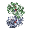
| ||||||||
|---|---|---|---|---|---|---|---|---|---|
| 1 |
| ||||||||
| Unit cell |
|
-Components
-Protein , 1 types, 2 molecules AB
| #1: Protein | Mass: 30918.793 Da / Num. of mol.: 2 Source method: isolated from a genetically manipulated source Source: (gene. exp.) Aminobacterium colombiense (bacteria) / Gene: Amico_1844 / Production host: |
|---|
-Non-polymers , 5 types, 338 molecules 


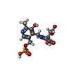

| #2: Chemical | ChemComp-EDO /  Mass: 62.068 Da / Num. of mol.: 4 / Source method: obtained synthetically / Formula: C2H6O2 Mass: 62.068 Da / Num. of mol.: 4 / Source method: obtained synthetically / Formula: C2H6O2#3: Chemical | ChemComp-NO3 /  Mass: 62.005 Da / Num. of mol.: 4 / Source method: obtained synthetically / Formula: NO3 Mass: 62.005 Da / Num. of mol.: 4 / Source method: obtained synthetically / Formula: NO3#4: Chemical |  Mass: 248.173 Da / Num. of mol.: 2 / Source method: obtained synthetically / Formula: C8H13N2O5P / Feature type: SUBJECT OF INVESTIGATION Mass: 248.173 Da / Num. of mol.: 2 / Source method: obtained synthetically / Formula: C8H13N2O5P / Feature type: SUBJECT OF INVESTIGATION#5: Chemical | ChemComp-PW0 / (~{ |  Mass: 376.256 Da / Num. of mol.: 1 / Source method: obtained synthetically / Formula: C13H17N2O9P / Feature type: SUBJECT OF INVESTIGATION Mass: 376.256 Da / Num. of mol.: 1 / Source method: obtained synthetically / Formula: C13H17N2O9P / Feature type: SUBJECT OF INVESTIGATION#6: Water | ChemComp-HOH / | Mass: 18.015 Da / Num. of mol.: 327 / Source method: isolated from a natural source / Formula: H2O |
|---|
-Details
| Has ligand of interest | Y |
|---|
-Experimental details
-Experiment
| Experiment | Method: X-RAY DIFFRACTION / Number of used crystals: 1 |
|---|
- Sample preparation
| Crystal | Density Matthews: 2.25 Å3/Da / Density % sol: 45.31 % |
|---|---|
| Crystal grow | Temperature: 288 K / Method: vapor diffusion, hanging drop / pH: 6.5 Details: 0.2M NaNitrate, 0.1M Bis-tris propane pH 6.5, 20% PEG3350 |
-Data collection
| Diffraction | Mean temperature: 100 K / Serial crystal experiment: N |
|---|---|
| Diffraction source | Source: ROTATING ANODE / Type: RIGAKU / Wavelength: 1.54184 Å |
| Detector | Type: RIGAKU HyPix-6000HE / Detector: PIXEL / Date: Jul 25, 2022 |
| Radiation | Protocol: SINGLE WAVELENGTH / Monochromatic (M) / Laue (L): M / Scattering type: x-ray |
| Radiation wavelength | Wavelength: 1.54184 Å / Relative weight: 1 |
| Reflection | Resolution: 1.85→19.76 Å / Num. obs: 48141 / % possible obs: 99.6 % / Redundancy: 11.6 % / CC1/2: 0.998 / Rmerge(I) obs: 0.113 / Rpim(I) all: 0.035 / Rrim(I) all: 0.119 / Χ2: 0.97 / Net I/σ(I): 15.9 / Num. measured all: 560284 |
| Reflection shell | Resolution: 1.85→1.89 Å / % possible obs: 99 % / Redundancy: 12.2 % / Rmerge(I) obs: 0.997 / Num. measured all: 35180 / Num. unique obs: 2886 / CC1/2: 0.832 / Rpim(I) all: 0.295 / Rrim(I) all: 1.041 / Χ2: 1 / Net I/σ(I) obs: 2.7 |
- Processing
| Software |
| ||||||||||||||||||||||||||||||||||||||||||||||||||||||||||||||||||||||||||||||||||||||||||||||||||||||||||||||||||||||||||||||||||||||||||||||||||||||||||||||||||||||||||||||||||||||
|---|---|---|---|---|---|---|---|---|---|---|---|---|---|---|---|---|---|---|---|---|---|---|---|---|---|---|---|---|---|---|---|---|---|---|---|---|---|---|---|---|---|---|---|---|---|---|---|---|---|---|---|---|---|---|---|---|---|---|---|---|---|---|---|---|---|---|---|---|---|---|---|---|---|---|---|---|---|---|---|---|---|---|---|---|---|---|---|---|---|---|---|---|---|---|---|---|---|---|---|---|---|---|---|---|---|---|---|---|---|---|---|---|---|---|---|---|---|---|---|---|---|---|---|---|---|---|---|---|---|---|---|---|---|---|---|---|---|---|---|---|---|---|---|---|---|---|---|---|---|---|---|---|---|---|---|---|---|---|---|---|---|---|---|---|---|---|---|---|---|---|---|---|---|---|---|---|---|---|---|---|---|---|---|
| Refinement | Method to determine structure: MOLECULAR REPLACEMENT / Resolution: 1.85→19.76 Å / Cor.coef. Fo:Fc: 0.951 / Cor.coef. Fo:Fc free: 0.918 / SU B: 3.947 / SU ML: 0.113 / Cross valid method: THROUGHOUT / ESU R: 0.156 / ESU R Free: 0.153 / Stereochemistry target values: MAXIMUM LIKELIHOOD / Details: HYDROGENS HAVE BEEN ADDED IN THE RIDING POSITIONS
| ||||||||||||||||||||||||||||||||||||||||||||||||||||||||||||||||||||||||||||||||||||||||||||||||||||||||||||||||||||||||||||||||||||||||||||||||||||||||||||||||||||||||||||||||||||||
| Solvent computation | Ion probe radii: 0.8 Å / Shrinkage radii: 0.8 Å / VDW probe radii: 1.2 Å / Solvent model: MASK | ||||||||||||||||||||||||||||||||||||||||||||||||||||||||||||||||||||||||||||||||||||||||||||||||||||||||||||||||||||||||||||||||||||||||||||||||||||||||||||||||||||||||||||||||||||||
| Displacement parameters | Biso mean: 21.494 Å2
| ||||||||||||||||||||||||||||||||||||||||||||||||||||||||||||||||||||||||||||||||||||||||||||||||||||||||||||||||||||||||||||||||||||||||||||||||||||||||||||||||||||||||||||||||||||||
| Refinement step | Cycle: 1 / Resolution: 1.85→19.76 Å
| ||||||||||||||||||||||||||||||||||||||||||||||||||||||||||||||||||||||||||||||||||||||||||||||||||||||||||||||||||||||||||||||||||||||||||||||||||||||||||||||||||||||||||||||||||||||
| Refine LS restraints |
|