 Movie
Movie Controller


+ Open data

- Basic information
| Entry | Database: PDB / ID: 8omj | ||||||
|---|---|---|---|---|---|---|---|
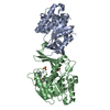
| Title | hKHK-C in complex with compound 28 | ||||||
 Components Components | Ketohexokinase | ||||||
 Keywords Keywords | SUGAR BINDING PROTEIN / KHK / Ketohexokinase / sugar binding kinase / inhibitor complex | ||||||
| Function / homology |  Function and homology information Function and homology informationEssential fructosuria / ketohexokinase / ketohexokinase activity / fructose binding / Fructose catabolism / regulation of glycogen metabolic process / response to sucrose / response to fructose / fructose metabolic process / response to zinc ion ...Essential fructosuria / ketohexokinase / ketohexokinase activity / fructose binding / Fructose catabolism / regulation of glycogen metabolic process / response to sucrose / response to fructose / fructose metabolic process / response to zinc ion / response to glucose / response to insulin / protein homodimerization activity / extracellular exosome / ATP binding / identical protein binding / cytoplasm / cytosol Similarity search - Function | ||||||
| Biological species |  Homo sapiens (human) Homo sapiens (human) | ||||||
| Method |  X-RAY DIFFRACTION / SYNCHROTRON / MOLECULAR REPLACEMENT / Resolution: 1.978 Å X-RAY DIFFRACTION / SYNCHROTRON / MOLECULAR REPLACEMENT / Resolution: 1.978 Å | ||||||
 Authors Authors | Pautsch, A. / Ebenhoch, R. | ||||||
| Funding support | 1items
| ||||||
 Citation Citation | Journal: Bioorg.Med.Chem.Lett. / Year: 2024 Title: Discovery of BI-9787, a potent zwitterionic ketohexokinase inhibitor with oral bioavailability. Authors: Heine, N. / Weber, A. / Pautsch, A. / Gottschling, D. / Uphues, I. / Bauer, M. / Ebenhoch, R. / Magarkar, A. / Nosse, B. / Kley, J.T. | ||||||
| History |
|
- Structure visualization
| Structure viewer | Molecule: MolmilJmol/JSmol |
|---|
- Downloads & links
-Download
| PDBx/mmCIF format | 8omj.cif.gz | 249.4 KB | Display | PDBx/mmCIF format |
|---|---|---|---|---|
| PDB format | pdb8omj.ent.gz | 200.8 KB | Display | PDB format |
| PDBx/mmJSON format | 8omj.json.gz | Tree view | PDBx/mmJSON format | |
| Others |  Other downloads Other downloads |
-Validation report
| Arichive directory | https://data.pdbj.org/pub/pdb/validation_reports/om/8omjftp://data.pdbj.org/pub/pdb/validation_reports/om/8omj | HTTPS FTP |
|---|
-Related structure data


-Links
 PDBj
PDBj- Assembly
| Deposited unit | 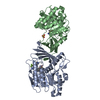
| ||||||||
|---|---|---|---|---|---|---|---|---|---|
| 1 |
| ||||||||
| Unit cell |
|
-Components
| #1: Protein | Mass: 34076.586 Da / Num. of mol.: 2 Source method: isolated from a genetically manipulated source Source: (gene. exp.) Homo sapiens (human) / Gene: KHK / Production host:  #2: Chemical | Mass: 527.500 Da / Num. of mol.: 2 / Source method: obtained synthetically / Formula: C22H25F3N5O3PS / Feature type: SUBJECT OF INVESTIGATION #3: Chemical |   Mass: 96.063 Da / Num. of mol.: 2 / Source method: obtained synthetically / Formula: SO4 Mass: 96.063 Da / Num. of mol.: 2 / Source method: obtained synthetically / Formula: SO4#4: Water | ChemComp-HOH / |  Mass: 18.015 Da / Num. of mol.: 398 / Source method: isolated from a natural source / Formula: H2O Mass: 18.015 Da / Num. of mol.: 398 / Source method: isolated from a natural source / Formula: H2OHas ligand of interest | Y | |
|---|
-Experimental details
-Experiment
| Experiment | Method: X-RAY DIFFRACTION / Number of used crystals: 1 |
|---|
- Sample preparation
| Crystal | Density Matthews: 3.46 Å3/Da / Density % sol: 64.48 % |
|---|---|
| Crystal grow | Temperature: 277 K / Method: vapor diffusion Details: 13% PEG 8000, 0.2 M ammonium sulfate, 0.1 M tri-sodium citrate pH 4.2 |
-Data collection
| Diffraction | Mean temperature: 100 K / Serial crystal experiment: N |
|---|---|
| Diffraction source | Source: SYNCHROTRON / Site: SLS  / Beamline: X06DA / Wavelength: 1 Å / Beamline: X06DA / Wavelength: 1 Å |
| Detector | Type: DECTRIS PILATUS 2M / Detector: PIXEL / Date: Feb 11, 2017 |
| Radiation | Protocol: SINGLE WAVELENGTH / Monochromatic (M) / Laue (L): M / Scattering type: x-ray |
| Radiation wavelength | Wavelength: 1 Å / Relative weight: 1 |
| Reflection | Resolution: 1.978→71.276 Å / Num. obs: 34330 / % possible obs: 93.5 % / Redundancy: 6.6 % / CC1/2: 0.997 / Rmerge(I) obs: 0.144 / Rpim(I) all: 0.06 / Net I/σ(I): 6.7 |
| Reflection shell | Resolution: 1.978→2.221 Å / Rmerge(I) obs: 1.062 / Mean I/σ(I) obs: 1.6 / Num. unique obs: 1716 / Rsym value: 1.062 / % possible all: 8.9 |
- Processing
| Software |
| |||||||||||||||||||||||||||||||||||||||||||||||||||||||||||||||||||||||||||
|---|---|---|---|---|---|---|---|---|---|---|---|---|---|---|---|---|---|---|---|---|---|---|---|---|---|---|---|---|---|---|---|---|---|---|---|---|---|---|---|---|---|---|---|---|---|---|---|---|---|---|---|---|---|---|---|---|---|---|---|---|---|---|---|---|---|---|---|---|---|---|---|---|---|---|---|---|
| Refinement | Method to determine structure: MOLECULAR REPLACEMENT / Resolution: 1.978→20.9 Å / Cor.coef. Fo:Fc: 0.93 / Cor.coef. Fo:Fc free: 0.896 / SU R Cruickshank DPI: 0.309 / Cross valid method: THROUGHOUT / SU R Blow DPI: 0.335 / SU Rfree Blow DPI: 0.243 / SU Rfree Cruickshank DPI: 0.238
| |||||||||||||||||||||||||||||||||||||||||||||||||||||||||||||||||||||||||||
| Displacement parameters | Biso mean: 44.34 Å2
| |||||||||||||||||||||||||||||||||||||||||||||||||||||||||||||||||||||||||||
| Refine analyze | Luzzati coordinate error obs: 0.31 Å | |||||||||||||||||||||||||||||||||||||||||||||||||||||||||||||||||||||||||||
| Refinement step | Cycle: LAST / Resolution: 1.978→20.9 Å
| |||||||||||||||||||||||||||||||||||||||||||||||||||||||||||||||||||||||||||
| Refine LS restraints |
| |||||||||||||||||||||||||||||||||||||||||||||||||||||||||||||||||||||||||||
| LS refinement shell | Resolution: 1.98→2.12 Å
| |||||||||||||||||||||||||||||||||||||||||||||||||||||||||||||||||||||||||||
| Refinement TLS params. | Refine-ID: X-RAY DIFFRACTION
| |||||||||||||||||||||||||||||||||||||||||||||||||||||||||||||||||||||||||||
| Refinement TLS group |
|
