[English] 日本語
Yorodumi
- PDB-8okf: WD repeat containing protein 5 (WDR5)- PER2 peptide -

+
Open data


ID or keywords:

Loading...

-
Basic information

Entry
Database: PDB / ID: 8okf
TitleWD repeat containing protein 5 (WDR5)- PER2 peptide
Components
  • Glutathione S-transferase class-mu 26 kDa isozyme,WD repeat domain 5
  • PER2 peptide
KeywordsCIRCADIAN CLOCK PROTEIN / WD40 repeat / peptide complex
Function / homology
Function and homology information


: / MLL3/4 complex / Set1C/COMPASS complex / ATAC complex / NSL complex / histone H3K4 methyltransferase activity / regulation of tubulin deacetylation / regulation of cell division / regulation of embryonic development / MLL1 complex ...: / MLL3/4 complex / Set1C/COMPASS complex / ATAC complex / NSL complex / histone H3K4 methyltransferase activity / regulation of tubulin deacetylation / regulation of cell division / regulation of embryonic development / MLL1 complex / glutathione transferase / glutathione transferase activity / positive regulation of gluconeogenesis / transcription initiation-coupled chromatin remodeling / methylated histone binding / skeletal system development / gluconeogenesis / regulation of cell cycle / negative regulation of transcription by RNA polymerase II
Similarity search - Function
Glutathione S-transferase, C-terminal domain / Glutathione S-transferase, N-terminal domain / Glutathione transferase family / Glutathione S-transferase, C-terminal / Soluble glutathione S-transferase N-terminal domain profile. / Glutathione S-transferase, C-terminal-like / Soluble glutathione S-transferase C-terminal domain profile. / Glutathione S-transferase, N-terminal / Glutathione S-transferase, C-terminal domain superfamily / Thioredoxin-like superfamily ...Glutathione S-transferase, C-terminal domain / Glutathione S-transferase, N-terminal domain / Glutathione transferase family / Glutathione S-transferase, C-terminal / Soluble glutathione S-transferase N-terminal domain profile. / Glutathione S-transferase, C-terminal-like / Soluble glutathione S-transferase C-terminal domain profile. / Glutathione S-transferase, N-terminal / Glutathione S-transferase, C-terminal domain superfamily / Thioredoxin-like superfamily / G-protein beta WD-40 repeat / WD40 repeat, conserved site / Trp-Asp (WD) repeats signature. / Trp-Asp (WD) repeats profile. / Trp-Asp (WD) repeats circular profile. / WD domain, G-beta repeat / WD40 repeats / WD40 repeat / WD40-repeat-containing domain superfamily / WD40/YVTN repeat-like-containing domain superfamily
Similarity search - Domain/homology
TRIS-HYDROXYMETHYL-METHYL-AMMONIUM / WD repeat domain 5 / Glutathione S-transferase class-mu 26 kDa isozyme
Similarity search - Component
Biological speciesMus musculus (house mouse)
MethodX-RAY DIFFRACTION / SYNCHROTRON / MOLECULAR REPLACEMENT / Resolution: 1.85 Å
AuthorsWolf, E. / Boergel, A.
Funding support Germany, 1items
OrganizationGrant numberCountry
Not funded Germany
CitationJournal: To Be Published
Title: A structural competition involving WDR5 times circadian oscillations
Authors: Wolf, E. / Boergel, A. / Rawleigh, A. / Conrady, M. / Hof, F. / Ricci, K. / Brown, S.
History
DepositionMar 28, 2023Deposition site: PDBE / Processing site: PDBE
Revision 1.0Apr 10, 2024Provider: repository / Type: Initial release

-
Structure visualization

Structure viewerMolecule:
MolmilJmol/JSmol

Downloads & links

-
Assembly

Deposited unit
A: Glutathione S-transferase class-mu 26 kDa isozyme,WD repeat domain 5
B: PER2 peptide
hetero molecules


Theoretical massNumber of molelcules
Total (without water)64,6947
Polymers64,1402
Non-polymers5535
Water1,76598
1


  • Idetical with deposited unit
  • defined by author
  • Evidence: gel filtration
TypeNameSymmetry operationNumber
identity operation1_555x,y,z1
Buried area2010 Å2
ΔGint-20 kcal/mol
Surface area11800 Å2
Unit cell
Length a, b, c (Å)78.152, 78.152, 101.748
Angle α, β, γ (deg.)90.00, 90.00, 90.00
Int Tables number94
Space group name H-MP42212

-
Components

-
Protein / Protein/peptide , 2 types, 2 molecules AB

#1: Protein Glutathione S-transferase class-mu 26 kDa isozyme,WD repeat domain 5 / GST 26 / Sj26 antigen / SjGST


Mass: 62673.555 Da / Num. of mol.: 1
Source method: isolated from a genetically manipulated source
Source: (gene. exp.) Mus musculus (house mouse) / Gene: WDR5 / Production host: Escherichia coli (E. coli)
References: UniProt: P08515, UniProt: A0A2K5BZZ6, glutathione transferase
#2: Protein/peptide PER2 peptide


Mass: 1466.656 Da / Num. of mol.: 1 / Source method: obtained synthetically / Source: (synth.) Mus musculus (house mouse)

-
Non-polymers , 4 types, 103 molecules

#3: Chemical ChemComp-144 / TRIS-HYDROXYMETHYL-METHYL-AMMONIUM


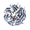
Mass: 122.143 Da / Num. of mol.: 2 / Source method: obtained synthetically / Formula: C4H12NO3
#4: Chemical ChemComp-CL / CHLORIDE ION / Chloride


Mass: 35.453 Da / Num. of mol.: 2 / Source method: obtained synthetically / Formula: Cl
#5: Chemical ChemComp-1PE / PENTAETHYLENE GLYCOL / PEG400 / Polyethylene glycol


Mass: 238.278 Da / Num. of mol.: 1 / Source method: obtained synthetically / Formula: C10H22O6 / Comment: precipitant*YM
#6: Water ChemComp-HOH / water / Water


Mass: 18.015 Da / Num. of mol.: 98 / Source method: isolated from a natural source / Formula: H2O

-
Details

Has ligand of interestN

-
Experimental details

-
Experiment

ExperimentMethod: X-RAY DIFFRACTION / Number of used crystals: 1

-
Sample preparation

Crystal growTemperature: 292 K / Method: vapor diffusion, hanging drop
Details: 0.1 M Tris pH 8.0, 50 mM sodium chloride, 13% PEG8000

-
Data collection

DiffractionMean temperature: 100 K / Serial crystal experiment: N
Diffraction sourceSource: SYNCHROTRON / Site: EMBL/DESY, HAMBURG / Beamline: X13 / Wavelength: 0.9763 Å
DetectorType: DECTRIS EIGER X 16M / Detector: PIXEL / Date: Nov 3, 2017
RadiationProtocol: SINGLE WAVELENGTH / Monochromatic (M) / Laue (L): M / Scattering type: x-ray
Radiation wavelengthWavelength: 0.9763 Å / Relative weight: 1
ReflectionResolution: 1.85→78.15 Å / Num. obs: 27636 / % possible obs: 100 % / Redundancy: 14.2 % / CC1/2: 0.999 / Net I/σ(I): 18.3
Reflection shellResolution: 1.85→1.89 Å / Num. unique obs: 1653 / CC1/2: 0.955

-
Processing

Software
NameVersionClassification
REFMAC5.8.0267refinement
XDSdata reduction
Aimlessdata scaling
PHASERphasing
RefinementMethod to determine structure: MOLECULAR REPLACEMENT / Resolution: 1.85→39.11 Å / Cor.coef. Fo:Fc: 0.958 / Cor.coef. Fo:Fc free: 0.939 / SU B: 2.383 / SU ML: 0.072 / Cross valid method: THROUGHOUT / ESU R: 0.123 / ESU R Free: 0.117 / Stereochemistry target values: MAXIMUM LIKELIHOOD / Details: HYDROGENS HAVE BEEN ADDED IN THE RIDING POSITIONS
RfactorNum. reflection% reflectionSelection details
Rfree0.2007 1336 4.8 %RANDOM
Rwork0.16513 ---
obs0.16684 26251 99.93 %-
Solvent computationIon probe radii: 0.8 Å / Shrinkage radii: 0.8 Å / VDW probe radii: 1.2 Å / Solvent model: MASK
Displacement parametersBiso mean: 18.936 Å2
Baniso -1Baniso -2Baniso -3
1--0.39 Å20 Å20 Å2
2---0.39 Å20 Å2
3---0.78 Å2
Refinement stepCycle: 1 / Resolution: 1.85→39.11 Å
ProteinNucleic acidLigandSolventTotal
Num. atoms2406 0 34 98 2538
Refine LS restraints
Refine-IDTypeDev idealDev ideal targetNumber
X-RAY DIFFRACTIONr_bond_refined_d0.0120.0132490
X-RAY DIFFRACTIONr_bond_other_d0.0010.0172330
X-RAY DIFFRACTIONr_angle_refined_deg1.6471.6293376
X-RAY DIFFRACTIONr_angle_other_deg1.4361.5865394
X-RAY DIFFRACTIONr_dihedral_angle_1_deg8.5485312
X-RAY DIFFRACTIONr_dihedral_angle_2_deg29.36324.84597
X-RAY DIFFRACTIONr_dihedral_angle_3_deg13.40215412
X-RAY DIFFRACTIONr_dihedral_angle_4_deg22.212153
X-RAY DIFFRACTIONr_chiral_restr0.080.2333
X-RAY DIFFRACTIONr_gen_planes_refined0.0090.022769
X-RAY DIFFRACTIONr_gen_planes_other0.0010.02529
X-RAY DIFFRACTIONr_nbd_refined
X-RAY DIFFRACTIONr_nbd_other
X-RAY DIFFRACTIONr_nbtor_refined
X-RAY DIFFRACTIONr_nbtor_other
X-RAY DIFFRACTIONr_xyhbond_nbd_refined
X-RAY DIFFRACTIONr_xyhbond_nbd_other
X-RAY DIFFRACTIONr_metal_ion_refined
X-RAY DIFFRACTIONr_metal_ion_other
X-RAY DIFFRACTIONr_symmetry_vdw_refined
X-RAY DIFFRACTIONr_symmetry_vdw_other
X-RAY DIFFRACTIONr_symmetry_hbond_refined
X-RAY DIFFRACTIONr_symmetry_hbond_other
X-RAY DIFFRACTIONr_symmetry_metal_ion_refined
X-RAY DIFFRACTIONr_symmetry_metal_ion_other
X-RAY DIFFRACTIONr_mcbond_it1.4761.8341254
X-RAY DIFFRACTIONr_mcbond_other1.4651.8321253
X-RAY DIFFRACTIONr_mcangle_it2.0542.741564
X-RAY DIFFRACTIONr_mcangle_other2.0592.7421565
X-RAY DIFFRACTIONr_scbond_it2.4082.0931235
X-RAY DIFFRACTIONr_scbond_other2.4072.0941236
X-RAY DIFFRACTIONr_scangle_it
X-RAY DIFFRACTIONr_scangle_other3.6233.0231813
X-RAY DIFFRACTIONr_long_range_B_refined4.43321.7462692
X-RAY DIFFRACTIONr_long_range_B_other4.43321.7582693
X-RAY DIFFRACTIONr_rigid_bond_restr
X-RAY DIFFRACTIONr_sphericity_free
X-RAY DIFFRACTIONr_sphericity_bonded
LS refinement shellResolution: 1.85→1.898 Å / Total num. of bins used: 20
RfactorNum. reflection% reflection
Rfree0.212 102 -
Rwork0.164 1900 -
obs--99.9 %

+
About Yorodumi

-
News

-
Feb 9, 2022. New format data for meta-information of EMDB entries

New format data for meta-information of EMDB entries

  • Version 3 of the EMDB header file is now the official format.
  • The previous official version 1.9 will be removed from the archive.

Related info.:EMDB header

External links:wwPDB to switch to version 3 of the EMDB data model

-
Aug 12, 2020. Covid-19 info

Covid-19 info

URL: https://pdbj.org/emnavi/covid19.php

New page: Covid-19 featured information page in EM Navigator.

Related info.:Covid-19 info / Mar 5, 2020. Novel coronavirus structure data

+
Mar 5, 2020. Novel coronavirus structure data

Novel coronavirus structure data

Related info.:Yorodumi Speices / Aug 12, 2020. Covid-19 info

External links:COVID-19 featured content - PDBj / Molecule of the Month (242):Coronavirus Proteases

+
Jan 31, 2019. EMDB accession codes are about to change! (news from PDBe EMDB page)

EMDB accession codes are about to change! (news from PDBe EMDB page)

  • The allocation of 4 digits for EMDB accession codes will soon come to an end. Whilst these codes will remain in use, new EMDB accession codes will include an additional digit and will expand incrementally as the available range of codes is exhausted. The current 4-digit format prefixed with “EMD-” (i.e. EMD-XXXX) will advance to a 5-digit format (i.e. EMD-XXXXX), and so on. It is currently estimated that the 4-digit codes will be depleted around Spring 2019, at which point the 5-digit format will come into force.
  • The EM Navigator/Yorodumi systems omit the EMD- prefix.

Related info.:Q: What is EMD? / ID/Accession-code notation in Yorodumi/EM Navigator

External links:EMDB Accession Codes are Changing Soon! / Contact to PDBj

+
Jul 12, 2017. Major update of PDB

Major update of PDB

  • wwPDB released updated PDB data conforming to the new PDBx/mmCIF dictionary.
  • This is a major update changing the version number from 4 to 5, and with Remediation, in which all the entries are updated.
  • In this update, many items about electron microscopy experimental information are reorganized (e.g. em_software).
  • Now, EM Navigator and Yorodumi are based on the updated data.

External links:wwPDB Remediation / Enriched Model Files Conforming to OneDep Data Standards Now Available in the PDB FTP Archive

-
Yorodumi

Thousand views of thousand structures

  • Yorodumi is a browser for structure data from EMDB, PDB, SASBDB, etc.
  • This page is also the successor to EM Navigator detail page, and also detail information page/front-end page for Omokage search.
  • The word "yorodu" (or yorozu) is an old Japanese word meaning "ten thousand". "mi" (miru) is to see.

Related info.:EMDB / PDB / SASBDB / Comparison of 3 databanks / Yorodumi Search / Aug 31, 2016. New EM Navigator & Yorodumi / Yorodumi Papers / Jmol/JSmol / Function and homology information / Changes in new EM Navigator and Yorodumi

Read more