 Movie
Movie Controller

[English] 日本語
 Yorodumi
- PDB-8ohy: Native Structure of Dictyostelium discoideum dye-decolorizing per... -
+ Open data

- Basic information
| Entry | Database: PDB / ID: 8ohy | ||||||
|---|---|---|---|---|---|---|---|
| Title | Native Structure of Dictyostelium discoideum dye-decolorizing peroxidase | ||||||
 Components Components | Peroxidase | ||||||
 Keywords Keywords | HYDROLASE / Dye-decolorizing peroxidase / H2O2-dependent oxidation / lignin degradation / hydrolysis / heme peroxidase | ||||||
| Function / homology |  Function and homology information Function and homology informationperoxidase activity / protein dimerization activity / heme binding / metal ion binding / cytoplasm / cytosol Similarity search - Function | ||||||
| Biological species |  | ||||||
| Method |  X-RAY DIFFRACTION / SYNCHROTRON / MOLECULAR REPLACEMENT / Resolution: 1.95 Å X-RAY DIFFRACTION / SYNCHROTRON / MOLECULAR REPLACEMENT / Resolution: 1.95 Å | ||||||
 Authors Authors | Koua, F.H. | ||||||
| Funding support | 1items
| ||||||
 Citation Citation | Journal: Front Chem / Year: 2023 Title: Heterologous expression, purification and structural features of native Dictyostelium discoideum dye-decolorizing peroxidase bound to a natively incorporated heme. Authors: Kalkan, O. / Kantamneni, S. / Brings, L. / Han, H. / Bean, R. / Mancuso, A.P. / Koua, F.H.M. | ||||||
| History |
|
- Structure visualization
| Structure viewer | Molecule: MolmilJmol/JSmol |
|---|
- Downloads & links
-Download
| PDBx/mmCIF format | 8ohy.cif.gz | 199.1 KB | Display | PDBx/mmCIF format |
|---|---|---|---|---|
| PDB format | pdb8ohy.ent.gz | 160.9 KB | Display | PDB format |
| PDBx/mmJSON format | 8ohy.json.gz | Tree view | PDBx/mmJSON format | |
| Others |  Other downloads Other downloads |
-Validation report
| Summary document | 8ohy_validation.pdf.gz | 809.8 KB | Display | wwPDB validaton report |
|---|---|---|---|---|
| Full document | 8ohy_full_validation.pdf.gz | 812.1 KB | Display | |
| Data in XML | 8ohy_validation.xml.gz | 14.7 KB | Display | |
| Data in CIF | 8ohy_validation.cif.gz | 20.5 KB | Display | |
| Arichive directory | https://data.pdbj.org/pub/pdb/validation_reports/oh/8ohyftp://data.pdbj.org/pub/pdb/validation_reports/oh/8ohy | HTTPS FTP |
-Related structure data
| Similar structure data |
|---|
-Links
 PDBj
PDBj- Assembly
| Deposited unit | 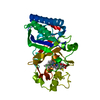
| ||||||||
|---|---|---|---|---|---|---|---|---|---|
| 1 | 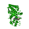
| ||||||||
| Unit cell |
|
-Components
| #1: Protein | Mass: 35014.855 Da / Num. of mol.: 1 Source method: isolated from a genetically manipulated source Source: (gene. exp.) Gene: DDB0168077, DDB0217308 / Production host:  |
|---|---|
| #2: Chemical | ChemComp-ZN /   Mass: 65.409 Da / Num. of mol.: 1 / Source method: obtained synthetically / Formula: Zn Mass: 65.409 Da / Num. of mol.: 1 / Source method: obtained synthetically / Formula: Zn |
| #3: Chemical | ChemComp-HEM /   Mass: 616.487 Da / Num. of mol.: 1 / Source method: isolated from a natural source / Formula: C34H32FeN4O4 / Feature type: SUBJECT OF INVESTIGATION Mass: 616.487 Da / Num. of mol.: 1 / Source method: isolated from a natural source / Formula: C34H32FeN4O4 / Feature type: SUBJECT OF INVESTIGATION |
| #4: Water | ChemComp-HOH /  Mass: 18.015 Da / Num. of mol.: 159 / Source method: isolated from a natural source / Formula: H2O Mass: 18.015 Da / Num. of mol.: 159 / Source method: isolated from a natural source / Formula: H2O |
| Has ligand of interest | Y |
-Experimental details
-Experiment
| Experiment | Method: X-RAY DIFFRACTION / Number of used crystals: 1 |
|---|
- Sample preparation
| Crystal | Density Matthews: 2.6 Å3/Da / Density % sol: 52.77 % |
|---|---|
| Crystal grow | Temperature: 293 K / Method: vapor diffusion, sitting drop / Details: HEPES-NaOH, PEG 6000, ZnCl |
-Data collection
| Diffraction | Mean temperature: 100 K / Serial crystal experiment: N |
|---|---|
| Diffraction source | Source: SYNCHROTRON / Site: PETRA III, DESY  / Beamline: P11 / Wavelength: 1.0332 Å / Beamline: P11 / Wavelength: 1.0332 Å |
| Detector | Type: DECTRIS EIGER X 16M / Detector: PIXEL / Date: Oct 1, 2022 |
| Radiation | Protocol: SINGLE WAVELENGTH / Monochromatic (M) / Laue (L): M / Scattering type: x-ray |
| Radiation wavelength | Wavelength: 1.0332 Å / Relative weight: 1 |
| Reflection | Resolution: 1.95→44.67 Å / Num. obs: 27351 / % possible obs: 98.54 % / Redundancy: 26.7 % / Biso Wilson estimate: 36.83 Å2 / CC1/2: 0.999 / CC star: 1 / Rmerge(I) obs: 0.2715 / Net I/σ(I): 12.02 |
| Reflection shell | Resolution: 1.95→2.02 Å / Redundancy: 26 % / Rmerge(I) obs: 4.643 / Mean I/σ(I) obs: 0.6 / Num. unique obs: 2472 / CC1/2: 0.411 / CC star: 0.764 / % possible all: 91.55 |
- Processing
| Software |
| |||||||||||||||||||||||||||||||||||||||||||||||||||||||||||||||
|---|---|---|---|---|---|---|---|---|---|---|---|---|---|---|---|---|---|---|---|---|---|---|---|---|---|---|---|---|---|---|---|---|---|---|---|---|---|---|---|---|---|---|---|---|---|---|---|---|---|---|---|---|---|---|---|---|---|---|---|---|---|---|---|---|
| Refinement | Method to determine structure: MOLECULAR REPLACEMENT / Resolution: 1.95→44.67 Å / SU ML: 0.3 / Cross valid method: FREE R-VALUE / σ(F): 1.33 / Phase error: 26.56 / Stereochemistry target values: ML
| |||||||||||||||||||||||||||||||||||||||||||||||||||||||||||||||
| Solvent computation | Shrinkage radii: 0.9 Å / VDW probe radii: 1.1 Å / Solvent model: FLAT BULK SOLVENT MODEL | |||||||||||||||||||||||||||||||||||||||||||||||||||||||||||||||
| Refinement step | Cycle: LAST / Resolution: 1.95→44.67 Å
| |||||||||||||||||||||||||||||||||||||||||||||||||||||||||||||||
| Refine LS restraints |
| |||||||||||||||||||||||||||||||||||||||||||||||||||||||||||||||
| LS refinement shell |
| |||||||||||||||||||||||||||||||||||||||||||||||||||||||||||||||
| Refinement TLS params. | Method: refined / Origin x: 2.0963 Å / Origin y: 23.1975 Å / Origin z: -17.3159 Å
| |||||||||||||||||||||||||||||||||||||||||||||||||||||||||||||||
| Refinement TLS group | Selection details: all |
