[English] 日本語
Yorodumi
- PDB-8og0: Crystal structure of MJF14-6-4-2 Fab fragment in complex with epi... -

+
Open data


ID or keywords:

Loading...

-
Basic information

Entry
Database: PDB / ID: 8og0
TitleCrystal structure of MJF14-6-4-2 Fab fragment in complex with epitope peptide
Components
  • Alpha-synuclein
  • Fab fragment heavy chain
  • Fab fragment light chain
KeywordsIMMUNE SYSTEM / Alpha synuclein / Fab fragment / MJF14-6-4-2 / epitope
Function / homology
Function and homology information


negative regulation of mitochondrial electron transport, NADH to ubiquinone / : / neutral lipid metabolic process / regulation of acyl-CoA biosynthetic process / negative regulation of dopamine uptake involved in synaptic transmission / negative regulation of norepinephrine uptake / positive regulation of SNARE complex assembly / positive regulation of hydrogen peroxide catabolic process / supramolecular fiber / mitochondrial membrane organization ...negative regulation of mitochondrial electron transport, NADH to ubiquinone / : / neutral lipid metabolic process / regulation of acyl-CoA biosynthetic process / negative regulation of dopamine uptake involved in synaptic transmission / negative regulation of norepinephrine uptake / positive regulation of SNARE complex assembly / positive regulation of hydrogen peroxide catabolic process / supramolecular fiber / mitochondrial membrane organization / negative regulation of chaperone-mediated autophagy / regulation of synaptic vesicle recycling / regulation of reactive oxygen species biosynthetic process / negative regulation of platelet-derived growth factor receptor signaling pathway / positive regulation of protein localization to cell periphery / negative regulation of exocytosis / regulation of glutamate secretion / response to iron(II) ion / SNARE complex assembly / positive regulation of neurotransmitter secretion / dopamine biosynthetic process / regulation of norepinephrine uptake / transporter regulator activity / regulation of locomotion / synaptic vesicle priming / mitochondrial ATP synthesis coupled electron transport / regulation of macrophage activation / positive regulation of inositol phosphate biosynthetic process / negative regulation of microtubule polymerization / synaptic vesicle transport / positive regulation of receptor recycling / dopamine uptake involved in synaptic transmission / protein kinase inhibitor activity / dynein complex binding / regulation of dopamine secretion / negative regulation of thrombin-activated receptor signaling pathway / cuprous ion binding / positive regulation of endocytosis / positive regulation of exocytosis / response to magnesium ion / synaptic vesicle exocytosis / enzyme inhibitor activity / kinesin binding / synaptic vesicle endocytosis / regulation of presynapse assembly / response to type II interferon / cysteine-type endopeptidase inhibitor activity / negative regulation of serotonin uptake / alpha-tubulin binding / supramolecular fiber organization / inclusion body / phospholipid metabolic process / cellular response to copper ion / axon terminus / cellular response to epinephrine stimulus / Hsp70 protein binding / response to interleukin-1 / regulation of microtubule cytoskeleton organization / SNARE binding / positive regulation of release of sequestered calcium ion into cytosol / adult locomotory behavior / negative regulation of protein kinase activity / excitatory postsynaptic potential / fatty acid metabolic process / phosphoprotein binding / protein tetramerization / microglial cell activation / regulation of long-term neuronal synaptic plasticity / synapse organization / ferrous iron binding / protein destabilization / PKR-mediated signaling / phospholipid binding / receptor internalization / tau protein binding / long-term synaptic potentiation / synaptic vesicle membrane / positive regulation of inflammatory response / actin cytoskeleton / actin binding / growth cone / cell cortex / cellular response to oxidative stress / neuron apoptotic process / chemical synaptic transmission / molecular adaptor activity / negative regulation of neuron apoptotic process / response to lipopolysaccharide / histone binding / amyloid fibril formation / lysosome / oxidoreductase activity / postsynapse / transcription cis-regulatory region binding / positive regulation of apoptotic process / Amyloid fiber formation / copper ion binding / response to xenobiotic stimulus / axon / neuronal cell body
Similarity search - Function
Synuclein / Alpha-synuclein / Synuclein
Similarity search - Domain/homology
Biological speciesOryctolagus cuniculus (rabbit)
Homo sapiens (human)
MethodX-RAY DIFFRACTION / SYNCHROTRON / MOLECULAR REPLACEMENT / Resolution: 1.712 Å
AuthorsTars, K. / Lieknina, I.
Funding supportEuropean Union, 1items
OrganizationGrant numberCountry
European Union (EU)OligoFITEuropean Union
CitationJournal: NPJ Parkinsons Dis / Year: 2024
Title: Structural basis of epitope recognition by anti-alpha-synuclein antibodies MJFR14-6-4-2.
Authors: Lieknina, I. / Reimer, L. / Pantelejevs, T. / Lends, A. / Jaudzems, K. / El-Turabi, A. / Gram, H. / Hammi, A. / Jensen, P.H. / Tars, K.
History
DepositionMar 17, 2023Deposition site: PDBE / Processing site: PDBE
Revision 1.0Mar 27, 2024Provider: repository / Type: Initial release
Revision 1.1Nov 6, 2024Group: Database references / Structure summary
Category: citation / citation_author ...citation / citation_author / pdbx_entry_details / pdbx_modification_feature
Item: _citation.journal_abbrev / _citation.journal_id_CSD ..._citation.journal_abbrev / _citation.journal_id_CSD / _citation.journal_id_ISSN / _citation.journal_volume / _citation.page_first / _citation.page_last / _citation.pdbx_database_id_DOI / _citation.pdbx_database_id_PubMed / _citation.title / _citation.year

-
Structure visualization

Structure viewerMolecule:
MolmilJmol/JSmol

Downloads & links

-
Assembly

Deposited unit
L: Fab fragment light chain
H: Fab fragment heavy chain
P: Alpha-synuclein


Theoretical massNumber of molelcules
Total (without water)46,8653
Polymers46,8653
Non-polymers00
Water3,081171
1


  • Idetical with deposited unit
  • defined by author
TypeNameSymmetry operationNumber
identity operation1_555x,y,z1
Buried area4490 Å2
ΔGint-31 kcal/mol
Surface area18250 Å2
Unit cell
Length a, b, c (Å)37.484, 66.700, 167.167
Angle α, β, γ (deg.)90.00, 90.00, 90.00
Int Tables number19
Space group name H-MP212121

-
Components

#1: Antibody Fab fragment light chain


Mass: 22771.078 Da / Num. of mol.: 1
Source method: isolated from a genetically manipulated source
Source: (gene. exp.) Oryctolagus cuniculus (rabbit) / Production host: Oryctolagus cuniculus (rabbit)
#2: Antibody Fab fragment heavy chain


Mass: 23486.291 Da / Num. of mol.: 1
Source method: isolated from a genetically manipulated source
Source: (gene. exp.) Oryctolagus cuniculus (rabbit) / Production host: Oryctolagus cuniculus (rabbit)
#3: Protein/peptide Alpha-synuclein / Non-A beta component of AD amyloid / Non-A4 component of amyloid precursor / NACP


Mass: 607.609 Da / Num. of mol.: 1 / Source method: obtained synthetically / Source: (synth.) Homo sapiens (human) / References: UniProt: P37840
#4: Water ChemComp-HOH / water


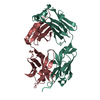
Mass: 18.015 Da / Num. of mol.: 171 / Source method: isolated from a natural source / Formula: H2O
Has protein modificationY

-
Experimental details

-
Experiment

ExperimentMethod: X-RAY DIFFRACTION / Number of used crystals: 1

-
Sample preparation

CrystalDensity Matthews: 2.28 Å3/Da / Density % sol: 46.05 %
Crystal growTemperature: 293 K / Method: vapor diffusion, sitting drop / pH: 5
Details: Sitting droplets were set up by mixing 1 ul of Fab fragments, 0.5 ul of peptide (1 mg/ml in 20 mM tris-HCl) and 1 ul crystallization buffer (24% PEG 6000, 0.1M Na-citrate pH 5.0).

-
Data collection

DiffractionMean temperature: 100 K / Serial crystal experiment: N
Diffraction sourceSource: SYNCHROTRON / Site: Diamond / Beamline: I03 / Wavelength: 0.976254 Å
DetectorType: DECTRIS EIGER2 XE 16M / Detector: PIXEL / Date: Feb 10, 2023
RadiationProtocol: SINGLE WAVELENGTH / Monochromatic (M) / Laue (L): M / Scattering type: x-ray
Radiation wavelengthWavelength: 0.976254 Å / Relative weight: 1
ReflectionResolution: 1.712→83.58 Å / Num. obs: 37216 / % possible obs: 94.3 % / Redundancy: 13 % / Rmerge(I) obs: 0.208 / Net I/σ(I): 8.5
Reflection shellResolution: 1.712→1.902 Å / Rmerge(I) obs: 1.609 / Num. unique obs: 1626

-
Processing

Software
NameVersionClassification
REFMAC5.8.0352refinement
autoPROCv1.0.5data reduction
autoPROCv1.0.5data scaling
PHASERv2.5.0phasing
RefinementMethod to determine structure: MOLECULAR REPLACEMENT / Resolution: 1.712→83.58 Å / Cor.coef. Fo:Fc: 0.952 / Cor.coef. Fo:Fc free: 0.936 / SU B: 3.525 / SU ML: 0.106 / Cross valid method: THROUGHOUT / ESU R: 0.171 / ESU R Free: 0.151 / Stereochemistry target values: MAXIMUM LIKELIHOOD / Details: HYDROGENS HAVE BEEN ADDED IN THE RIDING POSITIONS
RfactorNum. reflection% reflectionSelection details
Rfree0.23117 1594 4.9 %RANDOM
Rwork0.19531 ---
obs0.19704 30930 70.39 %-
Solvent computationIon probe radii: 0.8 Å / Shrinkage radii: 0.8 Å / VDW probe radii: 1.2 Å / Solvent model: MASK
Displacement parametersBiso mean: 22.835 Å2
Baniso -1Baniso -2Baniso -3
1--0.17 Å20 Å2-0 Å2
2--0.16 Å20 Å2
3---0.01 Å2
Refinement stepCycle: 1 / Resolution: 1.712→83.58 Å
ProteinNucleic acidLigandSolventTotal
Num. atoms3211 0 0 171 3382
Refine LS restraints
Refine-IDTypeDev idealDev ideal targetNumber
X-RAY DIFFRACTIONr_bond_refined_d0.0060.0113360
X-RAY DIFFRACTIONr_bond_other_d0.0010.0162927
X-RAY DIFFRACTIONr_angle_refined_deg1.3611.654617
X-RAY DIFFRACTIONr_angle_other_deg0.4771.5476848
X-RAY DIFFRACTIONr_dihedral_angle_1_deg7.6555445
X-RAY DIFFRACTIONr_dihedral_angle_2_deg6.766510
X-RAY DIFFRACTIONr_dihedral_angle_3_deg13.48710483
X-RAY DIFFRACTIONr_dihedral_angle_4_deg
X-RAY DIFFRACTIONr_chiral_restr0.0630.2542
X-RAY DIFFRACTIONr_gen_planes_refined0.0070.023862
X-RAY DIFFRACTIONr_gen_planes_other0.0010.02662
X-RAY DIFFRACTIONr_nbd_refined
X-RAY DIFFRACTIONr_nbd_other
X-RAY DIFFRACTIONr_nbtor_refined
X-RAY DIFFRACTIONr_nbtor_other
X-RAY DIFFRACTIONr_xyhbond_nbd_refined
X-RAY DIFFRACTIONr_xyhbond_nbd_other
X-RAY DIFFRACTIONr_metal_ion_refined
X-RAY DIFFRACTIONr_metal_ion_other
X-RAY DIFFRACTIONr_symmetry_vdw_refined
X-RAY DIFFRACTIONr_symmetry_vdw_other
X-RAY DIFFRACTIONr_symmetry_hbond_refined
X-RAY DIFFRACTIONr_symmetry_hbond_other
X-RAY DIFFRACTIONr_symmetry_metal_ion_refined
X-RAY DIFFRACTIONr_symmetry_metal_ion_other
X-RAY DIFFRACTIONr_mcbond_it1.9362.3991768
X-RAY DIFFRACTIONr_mcbond_other1.9352.3991768
X-RAY DIFFRACTIONr_mcangle_it2.9373.582217
X-RAY DIFFRACTIONr_mcangle_other2.9363.582218
X-RAY DIFFRACTIONr_scbond_it2.1892.4891592
X-RAY DIFFRACTIONr_scbond_other2.1882.4881593
X-RAY DIFFRACTIONr_scangle_it
X-RAY DIFFRACTIONr_scangle_other3.2663.6662401
X-RAY DIFFRACTIONr_long_range_B_refined4.70231.3753563
X-RAY DIFFRACTIONr_long_range_B_other4.65130.933540
X-RAY DIFFRACTIONr_rigid_bond_restr
X-RAY DIFFRACTIONr_sphericity_free
X-RAY DIFFRACTIONr_sphericity_bonded
LS refinement shellResolution: 1.712→1.756 Å / Total num. of bins used: 20
RfactorNum. reflection% reflection
Rfree0.357 9 -
Rwork0.309 136 -
obs--4.37 %

+
About Yorodumi

-
News

-
Feb 9, 2022. New format data for meta-information of EMDB entries

New format data for meta-information of EMDB entries

  • Version 3 of the EMDB header file is now the official format.
  • The previous official version 1.9 will be removed from the archive.

Related info.:EMDB header

External links:wwPDB to switch to version 3 of the EMDB data model

-
Aug 12, 2020. Covid-19 info

Covid-19 info

URL: https://pdbj.org/emnavi/covid19.php

New page: Covid-19 featured information page in EM Navigator.

Related info.:Covid-19 info / Mar 5, 2020. Novel coronavirus structure data

+
Mar 5, 2020. Novel coronavirus structure data

Novel coronavirus structure data

Related info.:Yorodumi Speices / Aug 12, 2020. Covid-19 info

External links:COVID-19 featured content - PDBj / Molecule of the Month (242):Coronavirus Proteases

+
Jan 31, 2019. EMDB accession codes are about to change! (news from PDBe EMDB page)

EMDB accession codes are about to change! (news from PDBe EMDB page)

  • The allocation of 4 digits for EMDB accession codes will soon come to an end. Whilst these codes will remain in use, new EMDB accession codes will include an additional digit and will expand incrementally as the available range of codes is exhausted. The current 4-digit format prefixed with “EMD-” (i.e. EMD-XXXX) will advance to a 5-digit format (i.e. EMD-XXXXX), and so on. It is currently estimated that the 4-digit codes will be depleted around Spring 2019, at which point the 5-digit format will come into force.
  • The EM Navigator/Yorodumi systems omit the EMD- prefix.

Related info.:Q: What is EMD? / ID/Accession-code notation in Yorodumi/EM Navigator

External links:EMDB Accession Codes are Changing Soon! / Contact to PDBj

+
Jul 12, 2017. Major update of PDB

Major update of PDB

  • wwPDB released updated PDB data conforming to the new PDBx/mmCIF dictionary.
  • This is a major update changing the version number from 4 to 5, and with Remediation, in which all the entries are updated.
  • In this update, many items about electron microscopy experimental information are reorganized (e.g. em_software).
  • Now, EM Navigator and Yorodumi are based on the updated data.

External links:wwPDB Remediation / Enriched Model Files Conforming to OneDep Data Standards Now Available in the PDB FTP Archive

-
Yorodumi

Thousand views of thousand structures

  • Yorodumi is a browser for structure data from EMDB, PDB, SASBDB, etc.
  • This page is also the successor to EM Navigator detail page, and also detail information page/front-end page for Omokage search.
  • The word "yorodu" (or yorozu) is an old Japanese word meaning "ten thousand". "mi" (miru) is to see.

Related info.:EMDB / PDB / SASBDB / Comparison of 3 databanks / Yorodumi Search / Aug 31, 2016. New EM Navigator & Yorodumi / Yorodumi Papers / Jmol/JSmol / Function and homology information / Changes in new EM Navigator and Yorodumi

Read more