 Movie
Movie Controller

[English] 日本語
 Yorodumi
- PDB-8ofd: Crystal structure of beta-conglutin from Lupinus albus refined to... -
+ Open data

- Basic information
| Entry | Database: PDB / ID: 8ofd | ||||||
|---|---|---|---|---|---|---|---|
| Title | Crystal structure of beta-conglutin from Lupinus albus refined to 2.81 A | ||||||
 Components Components | Conglutin beta 1 | ||||||
 Keywords Keywords | ALLERGEN / beta-conglutin / Lup an 1 / seed storage protein | ||||||
| Function / homology |  Function and homology information Function and homology information | ||||||
| Biological species |  Lupinus albus (white lupine) Lupinus albus (white lupine) | ||||||
| Method |  X-RAY DIFFRACTION / MOLECULAR REPLACEMENT / Resolution: 2.81 Å X-RAY DIFFRACTION / MOLECULAR REPLACEMENT / Resolution: 2.81 Å | ||||||
 Authors Authors | Dolot, R.M. / O'Sullivan, C.K. / Jauset-Rubio, M. | ||||||
| Funding support | 1items
| ||||||
 Citation Citation | Journal: To Be Published Title: First crystal structure of beta-conglutin, a major lupin allergen from Lupinus albus seeds Authors: Dolot, R.M. / O'Sullivan, C.K. / Jauset-Rubio, M. | ||||||
| History |
|
- Structure visualization
| Structure viewer | Molecule: MolmilJmol/JSmol |
|---|
- Downloads & links
-Download
| PDBx/mmCIF format | 8ofd.cif.gz | 102.4 KB | Display | PDBx/mmCIF format |
|---|---|---|---|---|
| PDB format | pdb8ofd.ent.gz | 74.4 KB | Display | PDB format |
| PDBx/mmJSON format | 8ofd.json.gz | Tree view | PDBx/mmJSON format | |
| Others |  Other downloads Other downloads |
-Validation report
| Summary document | 8ofd_validation.pdf.gz | 443.1 KB | Display | wwPDB validaton report |
|---|---|---|---|---|
| Full document | 8ofd_full_validation.pdf.gz | 455.7 KB | Display | |
| Data in XML | 8ofd_validation.xml.gz | 18.1 KB | Display | |
| Data in CIF | 8ofd_validation.cif.gz | 24.3 KB | Display | |
| Arichive directory | https://data.pdbj.org/pub/pdb/validation_reports/of/8ofdftp://data.pdbj.org/pub/pdb/validation_reports/of/8ofd | HTTPS FTP |
-Related structure data
| Similar structure data |
|---|
-Links
 PDBj
PDBj- Assembly
| Deposited unit | 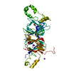
| ||||||||
|---|---|---|---|---|---|---|---|---|---|
| 1 | 
| ||||||||
| Unit cell |
| ||||||||
| Components on special symmetry positions |
|
-Components
| #1: Protein | Mass: 49029.992 Da / Num. of mol.: 1 / Source method: isolated from a natural source Details: 1-30 - signal protein (not present) 31-108 - propeptide (not present) 109-531 - conglutin beta 1. Residues 408-415 and 518-531 not included. Residue 176 changed from L to I based on experimental data. Source: (natural) Lupinus albus (white lupine) / References: UniProt: Q53HY0 |
|---|---|
| #2: Chemical | ChemComp-ACT /   Mass: 59.044 Da / Num. of mol.: 1 / Source method: obtained synthetically / Formula: C2H3O2 Mass: 59.044 Da / Num. of mol.: 1 / Source method: obtained synthetically / Formula: C2H3O2 |
| #3: Chemical | ChemComp-NA /   Mass: 22.990 Da / Num. of mol.: 1 / Source method: isolated from a natural source / Formula: Na Mass: 22.990 Da / Num. of mol.: 1 / Source method: isolated from a natural source / Formula: Na |
| #4: Chemical | ChemComp-K /   Mass: 39.098 Da / Num. of mol.: 1 / Source method: obtained synthetically / Formula: K Mass: 39.098 Da / Num. of mol.: 1 / Source method: obtained synthetically / Formula: K |
| #5: Water | ChemComp-HOH /  Mass: 18.015 Da / Num. of mol.: 82 / Source method: isolated from a natural source / Formula: H2O Mass: 18.015 Da / Num. of mol.: 82 / Source method: isolated from a natural source / Formula: H2O |
| Has ligand of interest | N |
-Experimental details
-Experiment
| Experiment | Method: X-RAY DIFFRACTION / Number of used crystals: 1 |
|---|
- Sample preparation
| Crystal | Density Matthews: 3.38 Å3/Da / Density % sol: 63.68 % |
|---|---|
| Crystal grow | Temperature: 281 K / Method: vapor diffusion, hanging drop / pH: 7 Details: 40% (v/v) Tacsimate pH 7.0, 50 mM HEPES pH 7.0, 2 mM Spermine, and 2 mM Hexaamine cobalt (III) |
-Data collection
| Diffraction | Mean temperature: 100 K / Serial crystal experiment: N |
|---|---|
| Diffraction source | Source: SEALED TUBE / Type: RIGAKU PhotonJet-S / Wavelength: 1.54184 Å |
| Detector | Type: RIGAKU HyPix-6000HE / Detector: PIXEL / Date: Apr 14, 2022 |
| Radiation | Protocol: SINGLE WAVELENGTH / Monochromatic (M) / Laue (L): M / Scattering type: x-ray |
| Radiation wavelength | Wavelength: 1.54184 Å / Relative weight: 1 |
| Reflection | Resolution: 2.81→21.77 Å / Num. obs: 15700 / % possible obs: 99.7 % / Redundancy: 7.8 % / Biso Wilson estimate: 55 Å2 / CC1/2: 0.993 / Rmerge(I) obs: 0.17 / Rpim(I) all: 0.064 / Rrim(I) all: 0.182 / Χ2: 0.93 / Net I/σ(I): 9.1 |
| Reflection shell | Resolution: 2.81→2.96 Å / Redundancy: 8.4 % / Rmerge(I) obs: 1.001 / Mean I/σ(I) obs: 2 / Num. unique obs: 2263 / CC1/2: 0.657 / Rpim(I) all: 0.366 / Rrim(I) all: 1.067 / Χ2: 0.92 / % possible all: 100 |
- Processing
| Software |
| ||||||||||||||||||||||||||||||||||||||||||||||||||||||||||||||||||||||||||||||||||||||||||||||||||||||||||||||||||||||||||||||||||||||||||||||||||||||||||||||||
|---|---|---|---|---|---|---|---|---|---|---|---|---|---|---|---|---|---|---|---|---|---|---|---|---|---|---|---|---|---|---|---|---|---|---|---|---|---|---|---|---|---|---|---|---|---|---|---|---|---|---|---|---|---|---|---|---|---|---|---|---|---|---|---|---|---|---|---|---|---|---|---|---|---|---|---|---|---|---|---|---|---|---|---|---|---|---|---|---|---|---|---|---|---|---|---|---|---|---|---|---|---|---|---|---|---|---|---|---|---|---|---|---|---|---|---|---|---|---|---|---|---|---|---|---|---|---|---|---|---|---|---|---|---|---|---|---|---|---|---|---|---|---|---|---|---|---|---|---|---|---|---|---|---|---|---|---|---|---|---|---|---|
| Refinement | Method to determine structure: MOLECULAR REPLACEMENT / Resolution: 2.81→21.767 Å / Cor.coef. Fo:Fc: 0.911 / Cor.coef. Fo:Fc free: 0.844 / WRfactor Rfree: 0.258 / WRfactor Rwork: 0.192 / SU B: 14.27 / SU ML: 0.303 / Average fsc free: 0.9384 / Average fsc work: 0.9696 / Cross valid method: THROUGHOUT / ESU R: 0.195 / ESU R Free: 0.08 Details: Hydrogens have been added in their riding positions
| ||||||||||||||||||||||||||||||||||||||||||||||||||||||||||||||||||||||||||||||||||||||||||||||||||||||||||||||||||||||||||||||||||||||||||||||||||||||||||||||||
| Solvent computation | Ion probe radii: 0.8 Å / Shrinkage radii: 0.8 Å / VDW probe radii: 1.2 Å / Solvent model: BABINET MODEL PLUS MASK | ||||||||||||||||||||||||||||||||||||||||||||||||||||||||||||||||||||||||||||||||||||||||||||||||||||||||||||||||||||||||||||||||||||||||||||||||||||||||||||||||
| Displacement parameters | Biso mean: 62.202 Å2
| ||||||||||||||||||||||||||||||||||||||||||||||||||||||||||||||||||||||||||||||||||||||||||||||||||||||||||||||||||||||||||||||||||||||||||||||||||||||||||||||||
| Refinement step | Cycle: LAST / Resolution: 2.81→21.767 Å
| ||||||||||||||||||||||||||||||||||||||||||||||||||||||||||||||||||||||||||||||||||||||||||||||||||||||||||||||||||||||||||||||||||||||||||||||||||||||||||||||||
| Refine LS restraints |
| ||||||||||||||||||||||||||||||||||||||||||||||||||||||||||||||||||||||||||||||||||||||||||||||||||||||||||||||||||||||||||||||||||||||||||||||||||||||||||||||||
| LS refinement shell |
|
