 Movie
Movie Controller

[English] 日本語
 Yorodumi
- PDB-8of3: Structure of the apoform of ALDEHYDE DEHYDROGENASE 5F1 (ALDH5F1) ... -
+ Open data

- Basic information
| Entry | Database: PDB / ID: 8of3 | ||||||||||||
|---|---|---|---|---|---|---|---|---|---|---|---|---|---|
| Title | Structure of the apoform of ALDEHYDE DEHYDROGENASE 5F1 (ALDH5F1) from the moss Physcomitrium patens | ||||||||||||
 Components Components | Succinate-semialdehyde dehydrogenase | ||||||||||||
 Keywords Keywords | OXIDOREDUCTASE / NAD+ binding / Succinate-semialdehyde dehydrogenase / ALDH5 | ||||||||||||
| Function / homology |  Function and homology information Function and homology informationsuccinate-semialdehyde dehydrogenase (NAD+) / succinate-semialdehyde dehydrogenase (NAD+) activity / gamma-aminobutyric acid catabolic process / nucleotide binding / mitochondrion Similarity search - Function | ||||||||||||
| Biological species |  Physcomitrium patens (plant) Physcomitrium patens (plant) | ||||||||||||
| Method |  X-RAY DIFFRACTION / SYNCHROTRON / MOLECULAR REPLACEMENT / Resolution: 1.348 Å X-RAY DIFFRACTION / SYNCHROTRON / MOLECULAR REPLACEMENT / Resolution: 1.348 Å | ||||||||||||
 Authors Authors | Morera, S. / Kopecny, D. / Vigouroux, A. | ||||||||||||
| Funding support |  Czech Republic, 3items Czech Republic, 3items
| ||||||||||||
 Citation Citation | Journal: To Be Published Title: A study on abiotic stress responses of aldehyde dehydrogenase (ALDH) superfamilies in moss and barley focused on members linked to the GABA shunt pathway Authors: Kopecny, D.J. / Belicek, D. / Vigouroux, A. / Luptakova, E. / Koncitikova, R. / von Schwartzenberg, K. / Cavar Zeljkovic, S. / Supikova, K. / Gruz, J. / Valarik, M. / Bergougnoux, V. / ...Authors: Kopecny, D.J. / Belicek, D. / Vigouroux, A. / Luptakova, E. / Koncitikova, R. / von Schwartzenberg, K. / Cavar Zeljkovic, S. / Supikova, K. / Gruz, J. / Valarik, M. / Bergougnoux, V. / Kopecny, D. / Morera, S. / Kopecna, M. | ||||||||||||
| History |
|
- Structure visualization
| Structure viewer | Molecule: MolmilJmol/JSmol |
|---|
- Downloads & links
-Download
| PDBx/mmCIF format | 8of3.cif.gz | 583.3 KB | Display | PDBx/mmCIF format |
|---|---|---|---|---|
| PDB format | pdb8of3.ent.gz | 490.1 KB | Display | PDB format |
| PDBx/mmJSON format | 8of3.json.gz | Tree view | PDBx/mmJSON format | |
| Others |  Other downloads Other downloads |
-Validation report
| Summary document | 8of3_validation.pdf.gz | 992.1 KB | Display | wwPDB validaton report |
|---|---|---|---|---|
| Full document | 8of3_full_validation.pdf.gz | 990.6 KB | Display | |
| Data in XML | 8of3_validation.xml.gz | 48.4 KB | Display | |
| Data in CIF | 8of3_validation.cif.gz | 75 KB | Display | |
| Arichive directory | https://data.pdbj.org/pub/pdb/validation_reports/of/8of3ftp://data.pdbj.org/pub/pdb/validation_reports/of/8of3 | HTTPS FTP |
-Related structure data


-Links
 PDBj
PDBj

- Assembly
| Deposited unit | 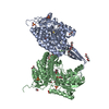
| ||||||||
|---|---|---|---|---|---|---|---|---|---|
| 1 | 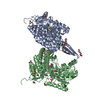
| ||||||||
| Unit cell |
|
-Components
-Protein , 1 types, 2 molecules AB
| #1: Protein | Mass: 54704.727 Da / Num. of mol.: 2 / Mutation: none Source method: isolated from a genetically manipulated source Source: (gene. exp.) Physcomitrium patens (plant) / Gene: PHYPA_030493Production host:  References: UniProt: A0A2K1ICJ7 |
|---|
-Non-polymers , 6 types, 1249 molecules 


| #2: Chemical |  Mass: 326.383 Da / Num. of mol.: 2 / Source method: obtained synthetically / Formula: C14H30O8 / Comment: precipitant*YM Mass: 326.383 Da / Num. of mol.: 2 / Source method: obtained synthetically / Formula: C14H30O8 / Comment: precipitant*YM#3: Chemical |  Mass: 150.173 Da / Num. of mol.: 3 / Source method: obtained synthetically / Formula: C6H14O4 Mass: 150.173 Da / Num. of mol.: 3 / Source method: obtained synthetically / Formula: C6H14O4#4: Chemical | ChemComp-1PE / |  Mass: 238.278 Da / Num. of mol.: 1 / Source method: obtained synthetically / Formula: C10H22O6 / Comment: precipitant*YM Mass: 238.278 Da / Num. of mol.: 1 / Source method: obtained synthetically / Formula: C10H22O6 / Comment: precipitant*YM#5: Chemical | ChemComp-EDO /  Mass: 62.068 Da / Num. of mol.: 9 / Source method: obtained synthetically / Formula: C2H6O2 Mass: 62.068 Da / Num. of mol.: 9 / Source method: obtained synthetically / Formula: C2H6O2#6: Chemical | ChemComp-SO4 /  Mass: 96.063 Da / Num. of mol.: 5 / Source method: obtained synthetically / Formula: SO4 Mass: 96.063 Da / Num. of mol.: 5 / Source method: obtained synthetically / Formula: SO4#7: Water | ChemComp-HOH / | Mass: 18.015 Da / Num. of mol.: 1229 / Source method: isolated from a natural source / Formula: H2O |
|---|
-Details
| Has ligand of interest | N |
|---|
-Experimental details
-Experiment
| Experiment | Method: X-RAY DIFFRACTION / Number of used crystals: 1 |
|---|
- Sample preparation
| Crystal | Density Matthews: 3.72 Å3/Da / Density % sol: 66.9 % |
|---|---|
| Crystal grow | Temperature: 292 K / Method: vapor diffusion / pH: 7.5 / Details: 1.7M LiSO4 |
-Data collection
| Diffraction | Mean temperature: 100 K / Serial crystal experiment: N |
|---|---|
| Diffraction source | Source: SYNCHROTRON / Site: SOLEIL  / Beamline: PROXIMA 2 / Wavelength: 0.99187 Å / Beamline: PROXIMA 2 / Wavelength: 0.99187 Å |
| Detector | Type: DECTRIS EIGER X 9M / Detector: PIXEL / Date: May 26, 2021 |
| Radiation | Protocol: SINGLE WAVELENGTH / Monochromatic (M) / Laue (L): M / Scattering type: x-ray |
| Radiation wavelength | Wavelength: 0.99187 Å / Relative weight: 1 |
| Reflection | Resolution: 1.34→131.14 Å / Num. obs: 241670 / % possible obs: 68 % / Redundancy: 20 % / CC1/2: 0.998 / Rmerge(I) obs: 0.156 / Net I/σ(I): 15.6 |
| Reflection shell | Resolution: 1.34→1.5 Å / Rmerge(I) obs: 1.8 / Num. unique obs: 12083 / CC1/2: 0.628 |
- Processing
| Software |
| ||||||||||||||||||||||||||||||||||||||||||||||||||||||||||||||||||||||||||||||||||||||||||||||||||||||||||||||||||
|---|---|---|---|---|---|---|---|---|---|---|---|---|---|---|---|---|---|---|---|---|---|---|---|---|---|---|---|---|---|---|---|---|---|---|---|---|---|---|---|---|---|---|---|---|---|---|---|---|---|---|---|---|---|---|---|---|---|---|---|---|---|---|---|---|---|---|---|---|---|---|---|---|---|---|---|---|---|---|---|---|---|---|---|---|---|---|---|---|---|---|---|---|---|---|---|---|---|---|---|---|---|---|---|---|---|---|---|---|---|---|---|---|---|---|---|
| Refinement | Method to determine structure: MOLECULAR REPLACEMENT / Resolution: 1.348→131.14 Å / Cor.coef. Fo:Fc: 0.971 / Cor.coef. Fo:Fc free: 0.966 / SU R Cruickshank DPI: 0.052 / Cross valid method: THROUGHOUT / σ(F): 0 / SU R Blow DPI: 0.05 / SU Rfree Blow DPI: 0.051 / SU Rfree Cruickshank DPI: 0.049 Details: HYDROGENS WERE FULLY REFINED WITH FULL OCCUPANCY AT NUCLEAR POSITION.
| ||||||||||||||||||||||||||||||||||||||||||||||||||||||||||||||||||||||||||||||||||||||||||||||||||||||||||||||||||
| Displacement parameters | Biso mean: 21 Å2
| ||||||||||||||||||||||||||||||||||||||||||||||||||||||||||||||||||||||||||||||||||||||||||||||||||||||||||||||||||
| Refine analyze | Luzzati coordinate error obs: 0.15 Å | ||||||||||||||||||||||||||||||||||||||||||||||||||||||||||||||||||||||||||||||||||||||||||||||||||||||||||||||||||
| Refinement step | Cycle: LAST / Resolution: 1.348→131.14 Å
| ||||||||||||||||||||||||||||||||||||||||||||||||||||||||||||||||||||||||||||||||||||||||||||||||||||||||||||||||||
| Refine LS restraints |
| ||||||||||||||||||||||||||||||||||||||||||||||||||||||||||||||||||||||||||||||||||||||||||||||||||||||||||||||||||
| LS refinement shell | Resolution: 1.35→1.44 Å / Total num. of bins used: 51
| ||||||||||||||||||||||||||||||||||||||||||||||||||||||||||||||||||||||||||||||||||||||||||||||||||||||||||||||||||
| Refinement TLS params. | Method: refined / Refine-ID: X-RAY DIFFRACTION
| ||||||||||||||||||||||||||||||||||||||||||||||||||||||||||||||||||||||||||||||||||||||||||||||||||||||||||||||||||
| Refinement TLS group |
|
