 Movie
Movie Controller

[English] 日本語
 Yorodumi
- PDB-8oez: Crystal structure of the Z-DNA hexamer d(CGCGCG) with Iron(II) ch... -
+ Open data

- Basic information
| Entry | Database: PDB / ID: 8oez | ||||||||||||||||||||||||||||
|---|---|---|---|---|---|---|---|---|---|---|---|---|---|---|---|---|---|---|---|---|---|---|---|---|---|---|---|---|---|
| Title | Crystal structure of the Z-DNA hexamer d(CGCGCG) with Iron(II) chloride | ||||||||||||||||||||||||||||
 Components Components | DNA (5'-D(* Keywords KeywordsDNA | Function / homology | : / DNA |  Function and homology information Function and homology informationBiological species | synthetic construct (others) | Method |  X-RAY DIFFRACTION / MOLECULAR REPLACEMENT / Resolution: 1.64 Å X-RAY DIFFRACTION / MOLECULAR REPLACEMENT / Resolution: 1.64 Å  Authors AuthorsLambert, M.C. / Hall, J.P. | Funding support | |  United Kingdom, 1items United Kingdom, 1items
 CitationJournal: To Be Published CitationJournal: To Be PublishedTitle: Iron binding preferences: observing metal coordination before oxidative damage. Authors: Lambert, M.C. / Hall, J.P. History |
|
- Structure visualization
| Structure viewer | Molecule: MolmilJmol/JSmol |
|---|
- Downloads & links
-Download
| PDBx/mmCIF format | 8oez.cif.gz | 32.1 KB | Display | PDBx/mmCIF format |
|---|---|---|---|---|
| PDB format | pdb8oez.ent.gz | 20.1 KB | Display | PDB format |
| PDBx/mmJSON format | 8oez.json.gz | Tree view | PDBx/mmJSON format | |
| Others |  Other downloads Other downloads |
-Validation report
| Arichive directory | https://data.pdbj.org/pub/pdb/validation_reports/oe/8oezftp://data.pdbj.org/pub/pdb/validation_reports/oe/8oez | HTTPS FTP |
|---|
-Related structure data
| Similar structure data |
|---|
-Links
 PDBj
PDBj


- Assembly
| Deposited unit | 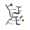
| |||||||||||||||||||||||||||
|---|---|---|---|---|---|---|---|---|---|---|---|---|---|---|---|---|---|---|---|---|---|---|---|---|---|---|---|---|
| 1 |
| |||||||||||||||||||||||||||
| Unit cell |
| |||||||||||||||||||||||||||
| Noncrystallographic symmetry (NCS) | NCS domain:
NCS domain segments:
NCS oper: (Code: givenMatrix: (0.209394902467, 0.977065595321, 0.0386858793464), (0.976591877247, -0.210957007194, 0.0420172156493), (0.0492146331497, 0.028982124761, -0.998367645874)Vector: -3. ...NCS oper: (Code: given Matrix: (0.209394902467, 0.977065595321, 0.0386858793464), Vector: |
-Components
| #1: DNA chain | Mass: 1810.205 Da / Num. of mol.: 2 / Source method: obtained synthetically / Source: (synth.) synthetic construct (others) #2: Chemical | ChemComp-FE2 /   Mass: 55.845 Da / Num. of mol.: 4 / Source method: obtained synthetically / Formula: Fe / Feature type: SUBJECT OF INVESTIGATION Mass: 55.845 Da / Num. of mol.: 4 / Source method: obtained synthetically / Formula: Fe / Feature type: SUBJECT OF INVESTIGATION#3: Chemical | ChemComp-CL / |   Mass: 35.453 Da / Num. of mol.: 1 / Source method: obtained synthetically / Formula: Cl Mass: 35.453 Da / Num. of mol.: 1 / Source method: obtained synthetically / Formula: Cl#4: Water | ChemComp-HOH / |  Mass: 18.015 Da / Num. of mol.: 52 / Source method: isolated from a natural source / Formula: H2O Mass: 18.015 Da / Num. of mol.: 52 / Source method: isolated from a natural source / Formula: H2OHas ligand of interest | Y | |
|---|
-Experimental details
-Experiment
| Experiment | Method: X-RAY DIFFRACTION / Number of used crystals: 1 |
|---|
- Sample preparation
| Crystal | Density Matthews: 1.61 Å3/Da / Density % sol: 23.78 % |
|---|---|
| Crystal grow | Temperature: 291 K / Method: vapor diffusion, sitting drop / pH: 6 Details: oligo, 2-methyl-2,4-pentanediol (MPD), sodium cacodylate, pH 6.0, KCl, NaCl and spermine tetrachloride |
-Data collection
| Diffraction | Mean temperature: 100 K / Serial crystal experiment: N |
|---|---|
| Diffraction source | Source: SEALED TUBE / Type: RIGAKU PhotonJet-S / Wavelength: 1.5418 Å |
| Detector | Type: RIGAKU HyPix-6000HE / Detector: PIXEL / Date: Oct 22, 2020 |
| Radiation | Protocol: SINGLE WAVELENGTH / Monochromatic (M) / Laue (L): M / Scattering type: x-ray |
| Radiation wavelength | Wavelength: 1.5418 Å / Relative weight: 1 |
| Reflection | Resolution: 1.64→14.6 Å / Num. obs: 3119 / % possible obs: 99.3 % / Redundancy: 2 % / Biso Wilson estimate: 6.55 Å2 / CC1/2: 1 / Rmerge(I) obs: 0.105 / Net I/σ(I): 13.7 |
| Reflection shell | Resolution: 1.64→1.7 Å / Rmerge(I) obs: 0.197 / Num. unique obs: 268 / CC1/2: 0.996 |
- Processing
| Software |
| ||||||||||||||||||||||||
|---|---|---|---|---|---|---|---|---|---|---|---|---|---|---|---|---|---|---|---|---|---|---|---|---|---|
| Refinement | Method to determine structure: MOLECULAR REPLACEMENT / Resolution: 1.64→14.6 Å / SU ML: 0.1678 / Cross valid method: FREE R-VALUE / σ(F): 1.42 / Phase error: 20.4767 Stereochemistry target values: GeoStd + Monomer Library + CDL v1.2
| ||||||||||||||||||||||||
| Solvent computation | Shrinkage radii: 0.9 Å / VDW probe radii: 1.1 Å / Solvent model: FLAT BULK SOLVENT MODEL | ||||||||||||||||||||||||
| Displacement parameters | Biso mean: 11.65 Å2 | ||||||||||||||||||||||||
| Refinement step | Cycle: LAST / Resolution: 1.64→14.6 Å
| ||||||||||||||||||||||||
| Refine LS restraints |
| ||||||||||||||||||||||||
| Refine LS restraints NCS | Type: Torsion NCS / Rms dev position: 0.414038578808 Å | ||||||||||||||||||||||||
| LS refinement shell |
|
