 Movie
Movie Controller


+ Open data

- Basic information
| Entry | Database: PDB / ID: 8kih | |||||||||
|---|---|---|---|---|---|---|---|---|---|---|
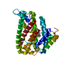
| Title | PhmA, a type I diterpene synthase without NST/DTE motif | |||||||||
 Components Components | diterpene synthase, PhmA | |||||||||
 Keywords Keywords | BIOSYNTHETIC PROTEIN / diterpene synthase | |||||||||
| Function / homology | Chem-FGG Function and homology information Function and homology information | |||||||||
| Biological species |  Phoma (fungus) Phoma (fungus) | |||||||||
| Method |  X-RAY DIFFRACTION / SYNCHROTRON / MOLECULAR REPLACEMENT / Resolution: 2 Å X-RAY DIFFRACTION / SYNCHROTRON / MOLECULAR REPLACEMENT / Resolution: 2 Å | |||||||||
 Authors Authors | Zhang, B. / Ge, H.M. / Zhu, A. / Zhang, Y. | |||||||||
| Funding support |  China, 2items China, 2items
| |||||||||
 Citation Citation | Journal: Angew.Chem.Int.Ed.Engl. / Year: 2023 Title: Biosynthesis of Phomactin Platelet Activating Factor Antagonist Requires a Two-Enzyme Cascade. Authors: Zhang, L. / Zhang, B. / Zhu, A. / Liu, S.H. / Wu, R. / Zhang, X. / Xu, Z. / Tan, R.X. / Ge, H.M. | |||||||||
| History |
|
- Structure visualization
| Structure viewer | Molecule: MolmilJmol/JSmol |
|---|
- Downloads & links
-Download
| PDBx/mmCIF format | 8kih.cif.gz | 171.3 KB | Display | PDBx/mmCIF format |
|---|---|---|---|---|
| PDB format | pdb8kih.ent.gz | 136.3 KB | Display | PDB format |
| PDBx/mmJSON format | 8kih.json.gz | Tree view | PDBx/mmJSON format | |
| Others |  Other downloads Other downloads |
-Validation report
| Arichive directory | https://data.pdbj.org/pub/pdb/validation_reports/ki/8kihftp://data.pdbj.org/pub/pdb/validation_reports/ki/8kih | HTTPS FTP |
|---|
-Related structure data
-Links
 PDBj
PDBj- Assembly
| Deposited unit | 
| |||||||||
|---|---|---|---|---|---|---|---|---|---|---|
| 1 |
| |||||||||
| Unit cell |
| |||||||||
| Components on special symmetry positions |
|
-Components
| #1: Protein | Mass: 37484.410 Da / Num. of mol.: 1 Source method: isolated from a genetically manipulated source Source: (gene. exp.) Phoma (fungus) / Strain: sp. ATCC 74077 / Production host:  | ||||||
|---|---|---|---|---|---|---|---|
| #2: Chemical | ChemComp-FGG / (  Mass: 468.434 Da / Num. of mol.: 1 / Source method: obtained synthetically / Formula: C20H35FO7P2 / Feature type: SUBJECT OF INVESTIGATION Mass: 468.434 Da / Num. of mol.: 1 / Source method: obtained synthetically / Formula: C20H35FO7P2 / Feature type: SUBJECT OF INVESTIGATION | ||||||
| #3: Chemical |   Mass: 24.305 Da / Num. of mol.: 2 / Source method: isolated from a natural source / Formula: Mg / Feature type: SUBJECT OF INVESTIGATION Mass: 24.305 Da / Num. of mol.: 2 / Source method: isolated from a natural source / Formula: Mg / Feature type: SUBJECT OF INVESTIGATION#4: Water | ChemComp-HOH / |  Mass: 18.015 Da / Num. of mol.: 229 / Source method: isolated from a natural source / Formula: H2O Mass: 18.015 Da / Num. of mol.: 229 / Source method: isolated from a natural source / Formula: H2OHas ligand of interest | Y | Has protein modification | N | |
-Experimental details
-Experiment
| Experiment | Method: X-RAY DIFFRACTION / Number of used crystals: 1 |
|---|
- Sample preparation
| Crystal | Density Matthews: 2.42 Å3/Da / Density % sol: 49.17 % |
|---|---|
| Crystal grow | Temperature: 291.15 K / Method: vapor diffusion / pH: 7.5 Details: 1.6 M Ammonium sulfate, 0.1 M Sodium chloride, 0.1 M Sodium HEPES pH 7.5 |
-Data collection
| Diffraction | Mean temperature: 100 K / Serial crystal experiment: N |
|---|---|
| Diffraction source | Source: SYNCHROTRON / Site: SSRF / Beamline: BL19U1 / Wavelength: 0.97851 Å |
| Detector | Type: DECTRIS PILATUS3 6M / Detector: PIXEL / Date: Dec 24, 2022 |
| Radiation | Protocol: MAD / Monochromatic (M) / Laue (L): M / Scattering type: x-ray |
| Radiation wavelength | Wavelength: 0.97851 Å / Relative weight: 1 |
| Reflection | Resolution: 1.8→35.44 Å / Num. obs: 33385 / % possible obs: 100 % / Redundancy: 19 % / CC1/2: 0.998 / Rmerge(I) obs: 0.121 / Rpim(I) all: 0.028 / Rrim(I) all: 0.124 / Χ2: 1.28 / Net I/σ(I): 17.4 / Num. measured all: 634671 |
| Reflection shell | Resolution: 1.8→1.84 Å / % possible obs: 100 % / Redundancy: 17.4 % / Rmerge(I) obs: 0.797 / Num. measured all: 34274 / Num. unique obs: 1975 / CC1/2: 0.876 / Rpim(I) all: 0.196 / Rrim(I) all: 0.821 / Χ2: 0.84 / Net I/σ(I) obs: 3.8 |
- Processing
| Software |
| |||||||||||||||||||||||||||||||||||||||||||||||||||||||||||||||||||||||||||||||||||||||||||||||||||||||||
|---|---|---|---|---|---|---|---|---|---|---|---|---|---|---|---|---|---|---|---|---|---|---|---|---|---|---|---|---|---|---|---|---|---|---|---|---|---|---|---|---|---|---|---|---|---|---|---|---|---|---|---|---|---|---|---|---|---|---|---|---|---|---|---|---|---|---|---|---|---|---|---|---|---|---|---|---|---|---|---|---|---|---|---|---|---|---|---|---|---|---|---|---|---|---|---|---|---|---|---|---|---|---|---|---|---|---|
| Refinement | Method to determine structure: MOLECULAR REPLACEMENT / Resolution: 2→34.88 Å / SU ML: 0.22 / Cross valid method: FREE R-VALUE / σ(F): 1.35 / Phase error: 22.24 / Stereochemistry target values: ML
| |||||||||||||||||||||||||||||||||||||||||||||||||||||||||||||||||||||||||||||||||||||||||||||||||||||||||
| Solvent computation | Shrinkage radii: 0.9 Å / VDW probe radii: 1.1 Å / Solvent model: FLAT BULK SOLVENT MODEL | |||||||||||||||||||||||||||||||||||||||||||||||||||||||||||||||||||||||||||||||||||||||||||||||||||||||||
| Refinement step | Cycle: LAST / Resolution: 2→34.88 Å
| |||||||||||||||||||||||||||||||||||||||||||||||||||||||||||||||||||||||||||||||||||||||||||||||||||||||||
| Refine LS restraints |
| |||||||||||||||||||||||||||||||||||||||||||||||||||||||||||||||||||||||||||||||||||||||||||||||||||||||||
| LS refinement shell |
| |||||||||||||||||||||||||||||||||||||||||||||||||||||||||||||||||||||||||||||||||||||||||||||||||||||||||
| Refinement TLS params. | Method: refined / Origin x: -36.418 Å / Origin y: -10.7393 Å / Origin z: -8.9853 Å
| |||||||||||||||||||||||||||||||||||||||||||||||||||||||||||||||||||||||||||||||||||||||||||||||||||||||||
| Refinement TLS group | Selection details: all |
