 Movie
Movie Controller


+ Open data

- Basic information
| Entry | Database: PDB / ID: 8kif | |||||||||
|---|---|---|---|---|---|---|---|---|---|---|
| Title | The structure of MmaE with substrate | |||||||||
 Components Components | Putative dioxygenase | |||||||||
 Keywords Keywords | OXIDOREDUCTASE / MmaE / Fe/2OG enzymes | |||||||||
| Function / homology | : / : / TauD/TfdA-like domain / Taurine catabolism dioxygenase TauD, TfdA family / Taurine dioxygenase TauD-like superfamily / dioxygenase activity / : / : / Putative dioxygenase Function and homology information Function and homology information | |||||||||
| Biological species |  Mycobacterium marinum M (bacteria) Mycobacterium marinum M (bacteria) | |||||||||
| Method |  X-RAY DIFFRACTION / SYNCHROTRON / MOLECULAR REPLACEMENT / Resolution: 2.13 Å X-RAY DIFFRACTION / SYNCHROTRON / MOLECULAR REPLACEMENT / Resolution: 2.13 Å | |||||||||
 Authors Authors | Chen, J. / Zhou, J. | |||||||||
| Funding support |  China, 1items China, 1items
| |||||||||
 Citation Citation | Journal: Acs Catalysis / Year: 2024 Title: Variation in biosynthesis and metal-binding properties of isonitrile-containing peptides produced by Mycobacteria versus Streptomyces. Authors: Chen, T.Y. / Chen, J. / Ruszczycky, M.W. / Hilovsky, D. / Hostetler, T. / Liu, X. / Zhou, J. / Chang, W.C. | |||||||||
| History |
|
- Structure visualization
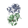
| Structure viewer | Molecule: MolmilJmol/JSmol |
|---|
- Downloads & links
-Download
| PDBx/mmCIF format | 8kif.cif.gz | 303.1 KB | Display | PDBx/mmCIF format |
|---|---|---|---|---|
| PDB format | pdb8kif.ent.gz | 197.2 KB | Display | PDB format |
| PDBx/mmJSON format | 8kif.json.gz | Tree view | PDBx/mmJSON format | |
| Others |  Other downloads Other downloads |
-Validation report
| Summary document | 8kif_validation.pdf.gz | 1.3 MB | Display | wwPDB validaton report |
|---|---|---|---|---|
| Full document | 8kif_full_validation.pdf.gz | 1.3 MB | Display | |
| Data in XML | 8kif_validation.xml.gz | 55.5 KB | Display | |
| Data in CIF | 8kif_validation.cif.gz | 73.3 KB | Display | |
| Arichive directory | https://data.pdbj.org/pub/pdb/validation_reports/ki/8kifftp://data.pdbj.org/pub/pdb/validation_reports/ki/8kif | HTTPS FTP |
-Related structure data
-Links
 PDBj
PDBj


- Assembly
| Deposited unit | 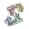
| ||||||||||||
|---|---|---|---|---|---|---|---|---|---|---|---|---|---|
| 1 | 
| ||||||||||||
| 2 | 
| ||||||||||||
| 3 | 
| ||||||||||||
| 4 | 
| ||||||||||||
| Unit cell |
|
-Components
| #1: Protein | Mass: 34107.648 Da / Num. of mol.: 4 Source method: isolated from a genetically manipulated source Source: (gene. exp.) Mycobacterium marinum M (bacteria) / Gene: MMRN_02610 / Production host: #2: Chemical | ChemComp-FE2 /   Mass: 55.845 Da / Num. of mol.: 4 / Source method: obtained synthetically / Formula: Fe Mass: 55.845 Da / Num. of mol.: 4 / Source method: obtained synthetically / Formula: Fe#3: Chemical | ChemComp-VY9 / ( Mass: 245.315 Da / Num. of mol.: 4 / Source method: obtained synthetically / Formula: C12H23NO4 / Feature type: SUBJECT OF INVESTIGATION #4: Water | ChemComp-HOH / |  Mass: 18.015 Da / Num. of mol.: 542 / Source method: isolated from a natural source / Formula: H2O Mass: 18.015 Da / Num. of mol.: 542 / Source method: isolated from a natural source / Formula: H2OHas ligand of interest | Y | Has protein modification | N | |
|---|
-Experimental details
-Experiment
| Experiment | Method: X-RAY DIFFRACTION / Number of used crystals: 1 |
|---|
- Sample preparation
| Crystal | Density Matthews: 2.49 Å3/Da / Density % sol: 50.67 % |
|---|---|
| Crystal grow | Temperature: 298 K / Method: vapor diffusion, sitting drop Details: 0.2 M Sodium acetate trihydrate, 0.1 M Tris/HCl (pH 8.5), 30% Polyethylene glycol 4000 |
-Data collection
| Diffraction | Mean temperature: 100 K / Serial crystal experiment: N |
|---|---|
| Diffraction source | Source: SYNCHROTRON / Site: SSRF / Beamline: BL17U1 / Wavelength: 0.9792 Å |
| Detector | Type: DECTRIS EIGER X 16M / Detector: PIXEL / Date: Nov 8, 2020 |
| Radiation | Protocol: SINGLE WAVELENGTH / Monochromatic (M) / Laue (L): M / Scattering type: x-ray |
| Radiation wavelength | Wavelength: 0.9792 Å / Relative weight: 1 |
| Reflection | Resolution: 2.13→61.98 Å / Num. obs: 65034 / % possible obs: 93.4 % / Redundancy: 3.6 % / Biso Wilson estimate: 31.74 Å2 / CC1/2: 0.997 / Net I/σ(I): 10.5 |
| Reflection shell | Resolution: 2.13→2.25 Å / Num. unique obs: 9227 / CC1/2: 0.851 |
- Processing
| Software |
| ||||||||||||||||||||||||||||||||||||||||||||||||||||||||||||||||||||||||||||||||||||||||||||||||||||||||||||||||||||||||||||||||||||||||||||||||||||||||||||||||||||||||
|---|---|---|---|---|---|---|---|---|---|---|---|---|---|---|---|---|---|---|---|---|---|---|---|---|---|---|---|---|---|---|---|---|---|---|---|---|---|---|---|---|---|---|---|---|---|---|---|---|---|---|---|---|---|---|---|---|---|---|---|---|---|---|---|---|---|---|---|---|---|---|---|---|---|---|---|---|---|---|---|---|---|---|---|---|---|---|---|---|---|---|---|---|---|---|---|---|---|---|---|---|---|---|---|---|---|---|---|---|---|---|---|---|---|---|---|---|---|---|---|---|---|---|---|---|---|---|---|---|---|---|---|---|---|---|---|---|---|---|---|---|---|---|---|---|---|---|---|---|---|---|---|---|---|---|---|---|---|---|---|---|---|---|---|---|---|---|---|---|---|
| Refinement | Method to determine structure: MOLECULAR REPLACEMENT / Resolution: 2.13→24.48 Å / SU ML: 0.256 / Cross valid method: FREE R-VALUE / σ(F): 1.97 / Phase error: 26.3736 / Stereochemistry target values: GeoStd + Monomer Library
| ||||||||||||||||||||||||||||||||||||||||||||||||||||||||||||||||||||||||||||||||||||||||||||||||||||||||||||||||||||||||||||||||||||||||||||||||||||||||||||||||||||||||
| Solvent computation | Shrinkage radii: 0.9 Å / VDW probe radii: 1.11 Å / Solvent model: FLAT BULK SOLVENT MODEL | ||||||||||||||||||||||||||||||||||||||||||||||||||||||||||||||||||||||||||||||||||||||||||||||||||||||||||||||||||||||||||||||||||||||||||||||||||||||||||||||||||||||||
| Displacement parameters | Biso mean: 39.05 Å2 | ||||||||||||||||||||||||||||||||||||||||||||||||||||||||||||||||||||||||||||||||||||||||||||||||||||||||||||||||||||||||||||||||||||||||||||||||||||||||||||||||||||||||
| Refinement step | Cycle: LAST / Resolution: 2.13→24.48 Å
| ||||||||||||||||||||||||||||||||||||||||||||||||||||||||||||||||||||||||||||||||||||||||||||||||||||||||||||||||||||||||||||||||||||||||||||||||||||||||||||||||||||||||
| Refine LS restraints |
| ||||||||||||||||||||||||||||||||||||||||||||||||||||||||||||||||||||||||||||||||||||||||||||||||||||||||||||||||||||||||||||||||||||||||||||||||||||||||||||||||||||||||
| LS refinement shell |
|
