[English] 日本語
Yorodumi
- PDB-8kg2: Crystal structure of p97-N/D1 hexamer in complex with FAF1-UBX domain -

+
Open data


ID or keywords:

Loading...

-
Basic information

Entry
Database: PDB / ID: 8kg2
TitleCrystal structure of p97-N/D1 hexamer in complex with FAF1-UBX domain
Components
  • FAS-associated factor 1
  • Transitional endoplasmic reticulum ATPase
KeywordsPROTEIN BINDING / valosin-containing protein / Fas-associated factor 1 / ubiquitin-regulatory X
Function / homology
Function and homology information


Fas signaling pathway / ooplasm / positive regulation of extrinsic apoptotic signaling pathway via death domain receptors / CD95 death-inducing signaling complex / : / flavin adenine dinucleotide catabolic process / VCP-NSFL1C complex / endosome to lysosome transport via multivesicular body sorting pathway / endoplasmic reticulum stress-induced pre-emptive quality control / BAT3 complex binding ...Fas signaling pathway / ooplasm / positive regulation of extrinsic apoptotic signaling pathway via death domain receptors / CD95 death-inducing signaling complex / : / flavin adenine dinucleotide catabolic process / VCP-NSFL1C complex / endosome to lysosome transport via multivesicular body sorting pathway / endoplasmic reticulum stress-induced pre-emptive quality control / BAT3 complex binding / cellular response to arsenite ion / protein-DNA covalent cross-linking repair / cytoplasm protein quality control / Derlin-1 retrotranslocation complex / positive regulation of protein K63-linked deubiquitination / positive regulation of oxidative phosphorylation / : / aggresome assembly / mitotic spindle disassembly / deubiquitinase activator activity / regulation of protein localization to chromatin / ubiquitin-modified protein reader activity / protein kinase regulator activity / VCP-NPL4-UFD1 AAA ATPase complex / cellular response to misfolded protein / negative regulation of protein localization to chromatin / vesicle-fusing ATPase / positive regulation of mitochondrial membrane potential / K48-linked polyubiquitin modification-dependent protein binding / regulation of aerobic respiration / retrograde protein transport, ER to cytosol / stress granule disassembly / regulation of synapse organization / ATPase complex / ubiquitin-specific protease binding / regulation of protein catabolic process / MHC class I protein binding / positive regulation of ATP biosynthetic process / ubiquitin-like protein ligase binding / RHOH GTPase cycle / polyubiquitin modification-dependent protein binding / autophagosome maturation / endoplasmic reticulum to Golgi vesicle-mediated transport / negative regulation of hippo signaling / NF-kappaB binding / HSF1 activation / translesion synthesis / interstrand cross-link repair / regulation of cell adhesion / proteasomal protein catabolic process / Protein methylation / ATP metabolic process / endoplasmic reticulum unfolded protein response / Attachment and Entry / Josephin domain DUBs / ERAD pathway / heat shock protein binding / lipid droplet / negative regulation of canonical NF-kappaB signal transduction / N-glycan trimming in the ER and Calnexin/Calreticulin cycle / proteasome complex / viral genome replication / positive regulation of DNA replication / Hh mutants are degraded by ERAD / ubiquitin binding / Hedgehog ligand biogenesis / Defective CFTR causes cystic fibrosis / Translesion Synthesis by POLH / negative regulation of smoothened signaling pathway / macroautophagy / positive regulation of protein-containing complex assembly / ABC-family proteins mediated transport / establishment of protein localization / positive regulation of non-canonical NF-kappaB signal transduction / ADP binding / autophagy / Aggrephagy / Ovarian tumor domain proteases / KEAP1-NFE2L2 pathway / cytoplasmic stress granule / positive regulation of protein catabolic process / azurophil granule lumen / positive regulation of canonical Wnt signaling pathway / E3 ubiquitin ligases ubiquitinate target proteins / nuclear envelope / positive regulation of proteasomal ubiquitin-dependent protein catabolic process / site of double-strand break / double-strand break repair / Neddylation / cellular response to heat / ubiquitin-dependent protein catabolic process / protein phosphatase binding / secretory granule lumen / regulation of apoptotic process / proteasome-mediated ubiquitin-dependent protein catabolic process / ficolin-1-rich granule lumen / Attachment and Entry / protein ubiquitination / ciliary basal body / positive regulation of apoptotic process
Similarity search - Function
FAS-associated factor 1-like, UBX domain / FAF1, UBA-like domain / : / Fas-associated factor 1 / UAS / : / UAS / Domain present in ubiquitin-regulatory proteins / UBX domain / UBX domain ...FAS-associated factor 1-like, UBX domain / FAF1, UBA-like domain / : / Fas-associated factor 1 / UAS / : / UAS / Domain present in ubiquitin-regulatory proteins / UBX domain / UBX domain / UBX domain profile. / UBA-like domain / AAA ATPase, CDC48 family / Cell division protein 48 (CDC48), N-terminal domain / CDC48, N-terminal subdomain / Cell division protein 48 (CDC48) N-terminal domain / CDC48, domain 2 / Cell division protein 48 (CDC48), domain 2 / Cell division protein 48 (CDC48) domain 2 / : / CDC48 domain 2-like superfamily / Aspartate decarboxylase-like domain superfamily / AAA ATPase, AAA+ lid domain / AAA+ lid domain / ATPase, AAA-type, conserved site / AAA-protein family signature. / ATPase family associated with various cellular activities (AAA) / ATPase, AAA-type, core / Thioredoxin-like superfamily / Ubiquitin-like domain superfamily / ATPases associated with a variety of cellular activities / AAA+ ATPase domain / P-loop containing nucleoside triphosphate hydrolase
Similarity search - Domain/homology
ADENOSINE-5'-DIPHOSPHATE / Transitional endoplasmic reticulum ATPase / FAS-associated factor 1
Similarity search - Component
Biological speciesHomo sapiens (human)
MethodX-RAY DIFFRACTION / SYNCHROTRON / MOLECULAR REPLACEMENT / Resolution: 3.1 Å
AuthorsKang, W.
Funding support Korea, Republic Of, 2items
OrganizationGrant numberCountry
National Research Foundation (NRF, Korea)2021R1A6A1A10044154 Korea, Republic Of
National Research Foundation (NRF, Korea)2022R1C1C1004221 Korea, Republic Of
CitationJournal: J. Korean Chem. Soc. / Year: 2024
Title: Crystal Structure of p97-N/D1 Hexamer Complexed with FAF1 UBX Domain
Authors: Kang, W.
History
DepositionAug 17, 2023Deposition site: PDBJ / Processing site: PDBJ
Revision 1.0May 29, 2024Provider: repository / Type: Initial release

-
Structure visualization

Structure viewerMolecule:
MolmilJmol/JSmol

Downloads & links

-
Assembly

Deposited unit
A: Transitional endoplasmic reticulum ATPase
B: Transitional endoplasmic reticulum ATPase
C: Transitional endoplasmic reticulum ATPase
D: Transitional endoplasmic reticulum ATPase
E: Transitional endoplasmic reticulum ATPase
F: Transitional endoplasmic reticulum ATPase
M: FAS-associated factor 1
N: FAS-associated factor 1
O: FAS-associated factor 1
P: FAS-associated factor 1
Q: FAS-associated factor 1
R: FAS-associated factor 1
G: Transitional endoplasmic reticulum ATPase
H: Transitional endoplasmic reticulum ATPase
I: Transitional endoplasmic reticulum ATPase
J: Transitional endoplasmic reticulum ATPase
K: Transitional endoplasmic reticulum ATPase
L: Transitional endoplasmic reticulum ATPase
S: FAS-associated factor 1
T: FAS-associated factor 1
U: FAS-associated factor 1
V: FAS-associated factor 1
W: FAS-associated factor 1
X: FAS-associated factor 1
hetero molecules


Theoretical massNumber of molelcules
Total (without water)699,47736
Polymers694,35124
Non-polymers5,12612
Water00
1


  • Idetical with deposited unit
  • defined by author
  • Evidence: gel filtration
TypeNameSymmetry operationNumber
identity operation1_555x,y,z1
Buried area72820 Å2
ΔGint-265 kcal/mol
Surface area238040 Å2
Unit cell
Length a, b, c (Å)106.665, 134.484, 148.223
Angle α, β, γ (deg.)71.52, 80.87, 87.53
Int Tables number1
Space group name H-MP1

-
Components

#1: Protein
Transitional endoplasmic reticulum ATPase


Mass: 48860.168 Da / Num. of mol.: 12 / Mutation: E192A, D193A, E194A
Source method: isolated from a genetically manipulated source
Source: (gene. exp.) Homo sapiens (human) / Gene: VCP / Production host: Escherichia coli (E. coli) / References: UniProt: P55072
#2: Protein
FAS-associated factor 1


Mass: 9002.397 Da / Num. of mol.: 12
Source method: isolated from a genetically manipulated source
Source: (gene. exp.) Homo sapiens (human) / Gene: FAF1 / Production host: Escherichia coli (E. coli) / References: UniProt: Q9UNN5
#3: Chemical
ChemComp-ADP / ADENOSINE-5'-DIPHOSPHATE


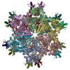
Mass: 427.201 Da / Num. of mol.: 12 / Source method: obtained synthetically / Formula: C10H15N5O10P2 / Feature type: SUBJECT OF INVESTIGATION / Comment: ADP, energy-carrying molecule*YM
Has ligand of interestY

-
Experimental details

-
Experiment

ExperimentMethod: X-RAY DIFFRACTION / Number of used crystals: 1

-
Sample preparation

CrystalDensity Matthews: 2.87 Å3/Da / Density % sol: 57.15 %
Crystal growTemperature: 295 K / Method: vapor diffusion, hanging drop / pH: 5.6
Details: 0.2M sodium acetate, 0.1M sodium citrate (pH 5.6), 5% polyethylene glycol 4000

-
Data collection

DiffractionMean temperature: 100 K / Serial crystal experiment: N
Diffraction sourceSource: SYNCHROTRON / Site: Photon Factory / Beamline: BL-17A / Wavelength: 1 Å
DetectorType: ADSC QUANTUM 210r / Detector: CCD / Date: Dec 19, 2009
RadiationProtocol: SINGLE WAVELENGTH / Monochromatic (M) / Laue (L): M / Scattering type: x-ray
Radiation wavelengthWavelength: 1 Å / Relative weight: 1
ReflectionResolution: 3.1→48.8 Å / Num. obs: 150419 / % possible obs: 97.4 % / Redundancy: 3.9 % / CC1/2: 0.998 / Rrim(I) all: 0.088 / Net I/σ(I): 7.7
Reflection shellResolution: 3.1→3.15 Å / Mean I/σ(I) obs: 1.2 / Num. unique obs: 7512 / CC1/2: 0.704 / Rrim(I) all: 0.839

-
Processing

Software
NameVersionClassification
REFMAC5.8.0419refinement
XDSdata reduction
Aimless1.12.12data scaling
PHASER2.8.3phasing
RefinementMethod to determine structure: MOLECULAR REPLACEMENT / Resolution: 3.1→48.8 Å / Cor.coef. Fo:Fc: 0.947 / Cor.coef. Fo:Fc free: 0.92 / SU B: 25.816 / SU ML: 0.429 / Cross valid method: THROUGHOUT / ESU R Free: 0.481 / Stereochemistry target values: MAXIMUM LIKELIHOOD
RfactorNum. reflection% reflectionSelection details
Rfree0.24626 6798 5 %RANDOM
Rwork0.19769 ---
obs0.20016 129500 97.36 %-
Solvent computationIon probe radii: 0.8 Å / Shrinkage radii: 0.8 Å / VDW probe radii: 1.2 Å / Solvent model: MASK
Displacement parametersBiso mean: 91.028 Å2
Baniso -1Baniso -2Baniso -3
1-3.91 Å22.83 Å21.86 Å2
2---0.14 Å21.64 Å2
3----1.48 Å2
Refinement stepCycle: 1 / Resolution: 3.1→48.8 Å
ProteinNucleic acidLigandSolventTotal
Num. atoms48411 0 324 0 48735
Refine LS restraints
Refine-IDTypeDev idealDev ideal targetNumber
X-RAY DIFFRACTIONr_bond_refined_d0.0060.01249565
X-RAY DIFFRACTIONr_bond_other_d
X-RAY DIFFRACTIONr_angle_refined_deg1.561.65267004
X-RAY DIFFRACTIONr_angle_other_deg
X-RAY DIFFRACTIONr_dihedral_angle_1_deg5.99356102
X-RAY DIFFRACTIONr_dihedral_angle_2_deg15.5055468
X-RAY DIFFRACTIONr_dihedral_angle_3_deg21.19109083
X-RAY DIFFRACTIONr_dihedral_angle_4_deg
X-RAY DIFFRACTIONr_chiral_restr0.1090.27631
X-RAY DIFFRACTIONr_gen_planes_refined0.0070.0236731
X-RAY DIFFRACTIONr_gen_planes_other
X-RAY DIFFRACTIONr_nbd_refined
X-RAY DIFFRACTIONr_nbd_other
X-RAY DIFFRACTIONr_nbtor_refined
X-RAY DIFFRACTIONr_nbtor_other
X-RAY DIFFRACTIONr_xyhbond_nbd_refined
X-RAY DIFFRACTIONr_xyhbond_nbd_other
X-RAY DIFFRACTIONr_metal_ion_refined
X-RAY DIFFRACTIONr_metal_ion_other
X-RAY DIFFRACTIONr_symmetry_vdw_refined
X-RAY DIFFRACTIONr_symmetry_vdw_other
X-RAY DIFFRACTIONr_symmetry_hbond_refined
X-RAY DIFFRACTIONr_symmetry_hbond_other
X-RAY DIFFRACTIONr_symmetry_metal_ion_refined
X-RAY DIFFRACTIONr_symmetry_metal_ion_other
X-RAY DIFFRACTIONr_mcbond_it9.6468.56224495
X-RAY DIFFRACTIONr_mcbond_other
X-RAY DIFFRACTIONr_mcangle_it14.99115.39130568
X-RAY DIFFRACTIONr_mcangle_other
X-RAY DIFFRACTIONr_scbond_it12.0899.42225070
X-RAY DIFFRACTIONr_scbond_other
X-RAY DIFFRACTIONr_scangle_it
X-RAY DIFFRACTIONr_scangle_other
X-RAY DIFFRACTIONr_long_range_B_refined25.19995.1873512
X-RAY DIFFRACTIONr_long_range_B_other
X-RAY DIFFRACTIONr_rigid_bond_restr
X-RAY DIFFRACTIONr_sphericity_free
X-RAY DIFFRACTIONr_sphericity_bonded
LS refinement shellResolution: 3.1→3.18 Å / Total num. of bins used: 20
RfactorNum. reflection% reflection
Rfree0.348 497 -
Rwork0.324 9608 -
obs--97.82 %

+
About Yorodumi

-
News

-
Feb 9, 2022. New format data for meta-information of EMDB entries

New format data for meta-information of EMDB entries

  • Version 3 of the EMDB header file is now the official format.
  • The previous official version 1.9 will be removed from the archive.

Related info.:EMDB header

External links:wwPDB to switch to version 3 of the EMDB data model

-
Aug 12, 2020. Covid-19 info

Covid-19 info

URL: https://pdbj.org/emnavi/covid19.php

New page: Covid-19 featured information page in EM Navigator.

Related info.:Covid-19 info / Mar 5, 2020. Novel coronavirus structure data

+
Mar 5, 2020. Novel coronavirus structure data

Novel coronavirus structure data

Related info.:Yorodumi Speices / Aug 12, 2020. Covid-19 info

External links:COVID-19 featured content - PDBj / Molecule of the Month (242):Coronavirus Proteases

+
Jan 31, 2019. EMDB accession codes are about to change! (news from PDBe EMDB page)

EMDB accession codes are about to change! (news from PDBe EMDB page)

  • The allocation of 4 digits for EMDB accession codes will soon come to an end. Whilst these codes will remain in use, new EMDB accession codes will include an additional digit and will expand incrementally as the available range of codes is exhausted. The current 4-digit format prefixed with “EMD-” (i.e. EMD-XXXX) will advance to a 5-digit format (i.e. EMD-XXXXX), and so on. It is currently estimated that the 4-digit codes will be depleted around Spring 2019, at which point the 5-digit format will come into force.
  • The EM Navigator/Yorodumi systems omit the EMD- prefix.

Related info.:Q: What is EMD? / ID/Accession-code notation in Yorodumi/EM Navigator

External links:EMDB Accession Codes are Changing Soon! / Contact to PDBj

+
Jul 12, 2017. Major update of PDB

Major update of PDB

  • wwPDB released updated PDB data conforming to the new PDBx/mmCIF dictionary.
  • This is a major update changing the version number from 4 to 5, and with Remediation, in which all the entries are updated.
  • In this update, many items about electron microscopy experimental information are reorganized (e.g. em_software).
  • Now, EM Navigator and Yorodumi are based on the updated data.

External links:wwPDB Remediation / Enriched Model Files Conforming to OneDep Data Standards Now Available in the PDB FTP Archive

-
Yorodumi

Thousand views of thousand structures

  • Yorodumi is a browser for structure data from EMDB, PDB, SASBDB, etc.
  • This page is also the successor to EM Navigator detail page, and also detail information page/front-end page for Omokage search.
  • The word "yorodu" (or yorozu) is an old Japanese word meaning "ten thousand". "mi" (miru) is to see.

Related info.:EMDB / PDB / SASBDB / Comparison of 3 databanks / Yorodumi Search / Aug 31, 2016. New EM Navigator & Yorodumi / Yorodumi Papers / Jmol/JSmol / Function and homology information / Changes in new EM Navigator and Yorodumi

Read more