 Movie
Movie Controller

[English] 日本語
 Yorodumi
- PDB-8kfw: Crystal structure of ZmMOC1 K229A in complex with a nicked Hollid... -
+ Open data

- Basic information
| Entry | Database: PDB / ID: 8kfw | ||||||
|---|---|---|---|---|---|---|---|
| Title | Crystal structure of ZmMOC1 K229A in complex with a nicked Holliday junction soaked in Mn2+ for 600 seconds | ||||||
 Components Components |
| ||||||
 Keywords Keywords | DNA BINDING PROTEIN / MOC1 / Holliday junction / Time-resolved crystallography | ||||||
| Function / homology | Holliday junction resolvase MOC1-like / crossover junction DNA endonuclease activity / metal ion binding / : / DNA / DNA (> 10) / Holliday junction resolvase MOC1, chloroplastic Function and homology information Function and homology information | ||||||
| Biological species |  | ||||||
| Method |  X-RAY DIFFRACTION / SYNCHROTRON / MOLECULAR REPLACEMENT / Resolution: 2.3 Å X-RAY DIFFRACTION / SYNCHROTRON / MOLECULAR REPLACEMENT / Resolution: 2.3 Å | ||||||
 Authors Authors | Zhang, D. / Luo, Z. / Lin, Z. | ||||||
| Funding support |  China, 1items China, 1items
| ||||||
 Citation Citation | Journal: Nat Commun / Year: 2024 Title: MOC1 cleaves Holliday junctions through a cooperative nick and counter-nick mechanism mediated by metal ions. Authors: Zhang, D. / Xu, S. / Luo, Z. / Lin, Z. | ||||||
| History |
|
- Structure visualization
| Structure viewer | Molecule: MolmilJmol/JSmol |
|---|
- Downloads & links
-Download
| PDBx/mmCIF format | 8kfw.cif.gz | 112.8 KB | Display | PDBx/mmCIF format |
|---|---|---|---|---|
| PDB format | pdb8kfw.ent.gz | 80.9 KB | Display | PDB format |
| PDBx/mmJSON format | 8kfw.json.gz | Tree view | PDBx/mmJSON format | |
| Others |  Other downloads Other downloads |
-Validation report
| Arichive directory | https://data.pdbj.org/pub/pdb/validation_reports/kf/8kfwftp://data.pdbj.org/pub/pdb/validation_reports/kf/8kfw | HTTPS FTP |
|---|
-Related structure data
| Related structure data |  8kfrC 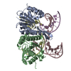 8kfsC 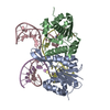 8kftC 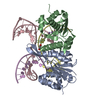 8kfuC 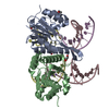 8kfvC 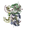 6jrgS S: Starting model for refinement C: citing same article ( |
|---|---|
| Similar structure data |
-Links
 PDBj
PDBj


- Assembly
| Deposited unit | 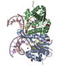
| ||||||||
|---|---|---|---|---|---|---|---|---|---|
| 1 |
| ||||||||
| Unit cell |
|
-Components
-Protein , 1 types, 2 molecules AB
| #1: Protein | Mass: 17558.018 Da / Num. of mol.: 2 / Mutation: K229A Source method: isolated from a genetically manipulated source Source: (gene. exp.)  |
|---|
-DNA chain , 3 types, 3 molecules CDE
| #2: DNA chain | Mass: 10137.505 Da / Num. of mol.: 1 / Source method: obtained synthetically / Source: (synth.) |
|---|---|
| #3: DNA chain | Mass: 7665.932 Da / Num. of mol.: 1 / Source method: obtained synthetically / Source: (synth.) |
| #4: DNA chain | Mass: 2426.617 Da / Num. of mol.: 1 / Source method: obtained synthetically / Source: (synth.) |
-Non-polymers , 3 types, 36 molecules 


| #5: Chemical | ChemComp-MN /  Mass: 54.938 Da / Num. of mol.: 6 / Source method: obtained synthetically / Formula: Mn Mass: 54.938 Da / Num. of mol.: 6 / Source method: obtained synthetically / Formula: Mn#6: Chemical | ChemComp-EDO /  Mass: 62.068 Da / Num. of mol.: 6 / Source method: obtained synthetically / Formula: C2H6O2 Mass: 62.068 Da / Num. of mol.: 6 / Source method: obtained synthetically / Formula: C2H6O2#7: Water | ChemComp-HOH / | Mass: 18.015 Da / Num. of mol.: 24 / Source method: isolated from a natural source / Formula: H2O |
|---|
-Details
| Has ligand of interest | N |
|---|
-Experimental details
-Experiment
| Experiment | Method: X-RAY DIFFRACTION / Number of used crystals: 1 |
|---|
- Sample preparation
| Crystal | Density Matthews: 2.44 Å3/Da / Density % sol: 49.6 % |
|---|---|
| Crystal grow | Temperature: 298 K / Method: vapor diffusion / pH: 6 / Details: 0.1 M MES pH 6.0, 35% Polyethylene glycol 400 |
-Data collection
| Diffraction | Mean temperature: 80 K / Serial crystal experiment: N |
|---|---|
| Diffraction source | Source: SYNCHROTRON / Site: SSRF / Beamline: BL18U1 / Wavelength: 1.5497 Å |
| Detector | Type: DECTRIS PILATUS 6M / Detector: PIXEL / Date: Nov 26, 2022 |
| Radiation | Protocol: SINGLE WAVELENGTH / Monochromatic (M) / Laue (L): M / Scattering type: x-ray |
| Radiation wavelength | Wavelength: 1.5497 Å / Relative weight: 1 |
| Reflection | Resolution: 2.3→49.07 Å / Num. obs: 23234 / % possible obs: 99.9 % / Redundancy: 6 % / CC1/2: 0.991 / Rmerge(I) obs: 0.123 / Net I/σ(I): 8.5 |
| Reflection shell | Resolution: 2.3→2.38 Å / Redundancy: 6.1 % / Rmerge(I) obs: 0.997 / Mean I/σ(I) obs: 1.7 / Num. unique obs: 2266 / CC1/2: 0.782 / % possible all: 100 |
- Processing
| Software |
| |||||||||||||||||||||||||||||||||||||||||||||||||||||||||||||||
|---|---|---|---|---|---|---|---|---|---|---|---|---|---|---|---|---|---|---|---|---|---|---|---|---|---|---|---|---|---|---|---|---|---|---|---|---|---|---|---|---|---|---|---|---|---|---|---|---|---|---|---|---|---|---|---|---|---|---|---|---|---|---|---|---|
| Refinement | Method to determine structure: MOLECULAR REPLACEMENT Starting model: 6JRG Resolution: 2.3→49.07 Å / SU ML: 0.29 / Cross valid method: THROUGHOUT / σ(F): 1.36 / Phase error: 26.62 / Stereochemistry target values: ML
| |||||||||||||||||||||||||||||||||||||||||||||||||||||||||||||||
| Solvent computation | Shrinkage radii: 0.9 Å / VDW probe radii: 1.1 Å / Solvent model: FLAT BULK SOLVENT MODEL | |||||||||||||||||||||||||||||||||||||||||||||||||||||||||||||||
| Refinement step | Cycle: LAST / Resolution: 2.3→49.07 Å
| |||||||||||||||||||||||||||||||||||||||||||||||||||||||||||||||
| Refine LS restraints |
| |||||||||||||||||||||||||||||||||||||||||||||||||||||||||||||||
| LS refinement shell |
|
