- PDB-8ke7: Crystal structure of DNA binding and cleavage core of human topoi... -
+
Open data
ID or keywords:
Loading...
-
Basic information
Entry
Database: PDB / ID: 8ke7
Title
Crystal structure of DNA binding and cleavage core of human topoisomerase 2-beta in a DNA binding-competent conformation
Components
DNA topoisomerase 2-beta
Keywords
DNA BINDING PROTEIN / eukaryotic DNA topoisomerase G-segment DNA binding cleavage center assembly
Function / homology
Function and homology information
positive regulation of single stranded viral RNA replication via double stranded DNA intermediate / sister chromatid segregation / resolution of meiotic recombination intermediates / DNA topoisomerase type II (double strand cut, ATP-hydrolyzing) activity / DNA topoisomerase (ATP-hydrolysing) / positive regulation of double-strand break repair via nonhomologous end joining / cellular response to ATP / forebrain development / DNA topological change / SUMOylation of DNA replication proteins ...positive regulation of single stranded viral RNA replication via double stranded DNA intermediate / sister chromatid segregation / resolution of meiotic recombination intermediates / DNA topoisomerase type II (double strand cut, ATP-hydrolyzing) activity / DNA topoisomerase (ATP-hydrolysing) / positive regulation of double-strand break repair via nonhomologous end joining / cellular response to ATP / forebrain development / DNA topological change / SUMOylation of DNA replication proteins / ribonucleoprotein complex binding / axonogenesis / B cell differentiation / cellular response to hydrogen peroxide / neuron migration / cellular senescence / ribonucleoprotein complex / chromatin binding / nucleolus / DNA binding / nucleoplasm / ATP binding / metal ion binding / nucleus / cytosol Similarity search - Function
DTHCT / DTHCT (NUC029) region / DNA topoisomerase 2, TOPRIM domain / C-terminal associated domain of TOPRIM / C-terminal associated domain of TOPRIM / DNA topoisomerase II, eukaryotic-type / : / Topoisomerase (Topo) IIA-type catalytic domain profile. / DNA topoisomerase, type IIA, alpha-helical domain superfamily / DNA topoisomerase, type IIA, domain A ...DTHCT / DTHCT (NUC029) region / DNA topoisomerase 2, TOPRIM domain / C-terminal associated domain of TOPRIM / C-terminal associated domain of TOPRIM / DNA topoisomerase II, eukaryotic-type / : / Topoisomerase (Topo) IIA-type catalytic domain profile. / DNA topoisomerase, type IIA, alpha-helical domain superfamily / DNA topoisomerase, type IIA, domain A / DNA topoisomerase, type IIA, domain A, alpha-beta / DNA gyrase/topoisomerase IV, subunit A / DNA Topoisomerase IV / DNA topoisomerase, type IIA, subunit B, domain 2 / DNA gyrase B / DNA topoisomerase, type IIA / DNA topoisomerase, type IIA, conserved site / DNA topoisomerase II signature. / TopoisomeraseII / DNA topoisomerase, type IIA, subunit B, C-terminal / Toprim domain / DNA topoisomerase, type IIA-like domain superfamily / Toprim domain profile. / TOPRIM domain / Histidine kinase-, DNA gyrase B-, and HSP90-like ATPase / Histidine kinase/HSP90-like ATPase superfamily / Ribosomal protein S5 domain 2-type fold, subgroup / Ribosomal protein S5 domain 2-type fold Similarity search - Domain/homology
 Movie
Movie Controller

 Yorodumi
 Open data
 Basic information
 Components
 Keywords
 Function and homology information
 Homo sapiens (human)
 X-RAY DIFFRACTION /
  Authors
 Taiwan, 1items
  Citation
 Structure visualization
 Downloads & links
 Other downloads
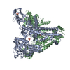
 PDBj
PDBj


 Assembly