 Movie
Movie Controller

[English] 日本語
 Yorodumi
- PDB-8kb2: Crystal Structure of M- and C-Domains of the shaft pilin LrpA fro... -
+ Open data

- Basic information
| Entry | Database: PDB / ID: 8kb2 | ||||||
|---|---|---|---|---|---|---|---|
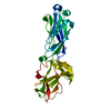
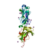
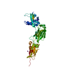
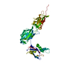
| Title | Crystal Structure of M- and C-Domains of the shaft pilin LrpA from Ligilactobacillus ruminis - iodide derivative | ||||||
 Components Components | LPXTG-motif cell wall anchor domain protein | ||||||
 Keywords Keywords | CELL ADHESION / Backbone pilin / isopeptide bond / pili / gut bacteria / probiotic | ||||||
| Function / homology |  Function and homology information Function and homology information: / Gram-positive pilin subunit D1, N-terminal / Gram-positive pilin subunit D1, N-terminal domain / Fimbrial isopeptide formation D2 domain / Prealbumin-like fold domain / Prealbumin-like fold domain / LPXTG cell wall anchor motif / Gram-positive cocci surface proteins LPxTG motif profile. / LPXTG cell wall anchor domain / Immunoglobulin-like fold Similarity search - Domain/homology | ||||||
| Biological species |  Ligilactobacillus ruminis ATCC 25644 (bacteria) Ligilactobacillus ruminis ATCC 25644 (bacteria) | ||||||
| Method |  X-RAY DIFFRACTION / SYNCHROTRON / SAD / Resolution: 1.91 Å X-RAY DIFFRACTION / SYNCHROTRON / SAD / Resolution: 1.91 Å | ||||||
 Authors Authors | Prajapati, A. / Palva, A. / von Ossowski, I. / Krishnan, V. | ||||||
| Funding support |  India, 1items India, 1items
| ||||||
 Citation Citation | Journal: Acta Crystallogr D Struct Biol / Year: 2024 Title: The crystal structure of the N-terminal domain of the backbone pilin LrpA reveals a new closure-and-twist motion for assembling dynamic pili in Ligilactobacillus ruminis. Authors: Prajapati, A. / Palva, A. / von Ossowski, I. / Krishnan, V. | ||||||
| History |
|
- Structure visualization
| Structure viewer | Molecule: MolmilJmol/JSmol |
|---|
- Downloads & links
-Download
| PDBx/mmCIF format | 8kb2.cif.gz | 119.3 KB | Display | PDBx/mmCIF format |
|---|---|---|---|---|
| PDB format | pdb8kb2.ent.gz | 90.5 KB | Display | PDB format |
| PDBx/mmJSON format | 8kb2.json.gz | Tree view | PDBx/mmJSON format | |
| Others |  Other downloads Other downloads |
-Validation report
| Arichive directory | https://data.pdbj.org/pub/pdb/validation_reports/kb/8kb2ftp://data.pdbj.org/pub/pdb/validation_reports/kb/8kb2 | HTTPS FTP |
|---|
-Related structure data
-Links
 PDBj
PDBj- Assembly
| Deposited unit | 
| ||||||||
|---|---|---|---|---|---|---|---|---|---|
| 1 |
| ||||||||
| Unit cell |
|
-Components
-Protein , 1 types, 1 molecules A
| #1: Protein | Mass: 28740.119 Da / Num. of mol.: 1 Source method: isolated from a genetically manipulated source Details: Shaft pilin Source: (gene. exp.) Ligilactobacillus ruminis ATCC 25644 (bacteria)Gene: HMPREF0542_11612 / Production host: |
|---|
-Non-polymers , 6 types, 166 molecules 


| #2: Chemical | ChemComp-IOD /  Mass: 126.904 Da / Num. of mol.: 13 / Source method: obtained synthetically / Formula: I / Feature type: SUBJECT OF INVESTIGATION Mass: 126.904 Da / Num. of mol.: 13 / Source method: obtained synthetically / Formula: I / Feature type: SUBJECT OF INVESTIGATION#3: Chemical |  Mass: 22.990 Da / Num. of mol.: 2 / Source method: obtained synthetically / Formula: Na / Feature type: SUBJECT OF INVESTIGATION Mass: 22.990 Da / Num. of mol.: 2 / Source method: obtained synthetically / Formula: Na / Feature type: SUBJECT OF INVESTIGATION#4: Chemical | ChemComp-CL / |  Mass: 35.453 Da / Num. of mol.: 1 / Source method: obtained synthetically / Formula: Cl / Feature type: SUBJECT OF INVESTIGATION Mass: 35.453 Da / Num. of mol.: 1 / Source method: obtained synthetically / Formula: Cl / Feature type: SUBJECT OF INVESTIGATION#5: Chemical | ChemComp-EDO / |  Mass: 62.068 Da / Num. of mol.: 1 / Source method: obtained synthetically / Formula: C2H6O2 / Feature type: SUBJECT OF INVESTIGATION Mass: 62.068 Da / Num. of mol.: 1 / Source method: obtained synthetically / Formula: C2H6O2 / Feature type: SUBJECT OF INVESTIGATION#6: Chemical |  Mass: 92.094 Da / Num. of mol.: 2 / Source method: obtained synthetically / Formula: C3H8O3 / Feature type: SUBJECT OF INVESTIGATION Mass: 92.094 Da / Num. of mol.: 2 / Source method: obtained synthetically / Formula: C3H8O3 / Feature type: SUBJECT OF INVESTIGATION#7: Water | ChemComp-HOH / | Mass: 18.015 Da / Num. of mol.: 147 / Source method: isolated from a natural source / Formula: H2O |
|---|
-Details
| Has ligand of interest | Y |
|---|
-Experimental details
-Experiment
| Experiment | Method: X-RAY DIFFRACTION / Number of used crystals: 1 |
|---|
- Sample preparation
| Crystal | Density Matthews: 2.33 Å3/Da / Density % sol: 47.22 % |
|---|---|
| Crystal grow | Temperature: 295 K / Method: vapor diffusion, hanging drop / pH: 4.6 / Details: 0.1 M sodium acetate pH 4.6, 25% PEG 4000 |
-Data collection
| Diffraction | Mean temperature: 100 K / Serial crystal experiment: N |
|---|---|
| Diffraction source | Source: SYNCHROTRON / Site: ELETTRA  / Beamline: 11.2C / Wavelength: 1.5498 Å / Beamline: 11.2C / Wavelength: 1.5498 Å |
| Detector | Type: DECTRIS PILATUS 6M / Detector: PIXEL / Date: Sep 6, 2019 |
| Radiation | Protocol: SINGLE WAVELENGTH / Monochromatic (M) / Laue (L): M / Scattering type: x-ray |
| Radiation wavelength | Wavelength: 1.5498 Å / Relative weight: 1 |
| Reflection | Resolution: 1.905→39.58 Å / Num. obs: 21167 / % possible obs: 97.6 % / Redundancy: 48.4 % / CC1/2: 0.999 / Rmerge(I) obs: 0.129 / Rpim(I) all: 0.018 / Rrim(I) all: 0.13 / Net I/σ(I): 24.2 |
| Reflection shell | Resolution: 1.905→1.945 Å / Redundancy: 45.5 % / Rmerge(I) obs: 0.97 / Mean I/σ(I) obs: 4.8 / Num. unique obs: 1058 / CC1/2: 0.969 / Rpim(I) all: 0.141 / % possible all: 85.5 |
- Processing
| Software |
| ||||||||||||||||||||||||||||||||||||||||||||||||||||||||||||||||||||||||||||||||||||||||||||||||||||||||||||||||||||||||||||||||||||||||||||||||||||||||||||||||||||||||||||||||||||||
|---|---|---|---|---|---|---|---|---|---|---|---|---|---|---|---|---|---|---|---|---|---|---|---|---|---|---|---|---|---|---|---|---|---|---|---|---|---|---|---|---|---|---|---|---|---|---|---|---|---|---|---|---|---|---|---|---|---|---|---|---|---|---|---|---|---|---|---|---|---|---|---|---|---|---|---|---|---|---|---|---|---|---|---|---|---|---|---|---|---|---|---|---|---|---|---|---|---|---|---|---|---|---|---|---|---|---|---|---|---|---|---|---|---|---|---|---|---|---|---|---|---|---|---|---|---|---|---|---|---|---|---|---|---|---|---|---|---|---|---|---|---|---|---|---|---|---|---|---|---|---|---|---|---|---|---|---|---|---|---|---|---|---|---|---|---|---|---|---|---|---|---|---|---|---|---|---|---|---|---|---|---|---|---|
| Refinement | Method to determine structure: SAD / Resolution: 1.91→39.58 Å / Cor.coef. Fo:Fc: 0.951 / Cor.coef. Fo:Fc free: 0.939 / SU B: 4.982 / SU ML: 0.08 / Cross valid method: THROUGHOUT / ESU R: 0.155 / ESU R Free: 0.137 / Stereochemistry target values: MAXIMUM LIKELIHOOD / Details: HYDROGENS HAVE BEEN ADDED IN THE RIDING POSITIONS
| ||||||||||||||||||||||||||||||||||||||||||||||||||||||||||||||||||||||||||||||||||||||||||||||||||||||||||||||||||||||||||||||||||||||||||||||||||||||||||||||||||||||||||||||||||||||
| Solvent computation | Ion probe radii: 0.8 Å / Shrinkage radii: 0.8 Å / VDW probe radii: 1.2 Å / Solvent model: MASK | ||||||||||||||||||||||||||||||||||||||||||||||||||||||||||||||||||||||||||||||||||||||||||||||||||||||||||||||||||||||||||||||||||||||||||||||||||||||||||||||||||||||||||||||||||||||
| Displacement parameters | Biso mean: 20.742 Å2
| ||||||||||||||||||||||||||||||||||||||||||||||||||||||||||||||||||||||||||||||||||||||||||||||||||||||||||||||||||||||||||||||||||||||||||||||||||||||||||||||||||||||||||||||||||||||
| Refinement step | Cycle: 1 / Resolution: 1.91→39.58 Å
| ||||||||||||||||||||||||||||||||||||||||||||||||||||||||||||||||||||||||||||||||||||||||||||||||||||||||||||||||||||||||||||||||||||||||||||||||||||||||||||||||||||||||||||||||||||||
| Refine LS restraints |
|