 Movie
Movie Controller


+ Open data

- Basic information
| Entry | Database: PDB / ID: 8k8l | ||||||
|---|---|---|---|---|---|---|---|
| Title | Structure of Klebsiella pneumonia ModA with molybdate | ||||||
 Components Components | Molybdate transporter periplasmic protein | ||||||
 Keywords Keywords | METAL BINDING PROTEIN / Molybdate binding protein | ||||||
| Function / homology |  Function and homology information Function and homology informationtungstate binding / molybdate ion binding / molybdate ion transport / outer membrane-bounded periplasmic space / metal ion binding Similarity search - Function | ||||||
| Biological species |  Klebsiella pneumoniae subsp. pneumoniae (bacteria) Klebsiella pneumoniae subsp. pneumoniae (bacteria) | ||||||
| Method |  X-RAY DIFFRACTION / SYNCHROTRON / MOLECULAR REPLACEMENT / Resolution: 1.77 Å X-RAY DIFFRACTION / SYNCHROTRON / MOLECULAR REPLACEMENT / Resolution: 1.77 Å | ||||||
 Authors Authors | Zhao, Q. / Bartlam, M. | ||||||
| Funding support |  China, 1items China, 1items
| ||||||
 Citation Citation | Journal: Biochem.Biophys.Res.Commun. / Year: 2023 Title: Structural analysis of molybdate binding protein ModA from Klebsiella pneumoniae. Authors: Zhao, Q. / Su, X. / Wang, Y. / Liu, R. / Bartlam, M. | ||||||
| History |
|
- Structure visualization
| Structure viewer | Molecule: MolmilJmol/JSmol |
|---|
- Downloads & links
-Download
| PDBx/mmCIF format | 8k8l.cif.gz | 65.7 KB | Display | PDBx/mmCIF format |
|---|---|---|---|---|
| PDB format | pdb8k8l.ent.gz | 44.8 KB | Display | PDB format |
| PDBx/mmJSON format | 8k8l.json.gz | Tree view | PDBx/mmJSON format | |
| Others |  Other downloads Other downloads |
-Validation report
| Arichive directory | https://data.pdbj.org/pub/pdb/validation_reports/k8/8k8lftp://data.pdbj.org/pub/pdb/validation_reports/k8/8k8l | HTTPS FTP |
|---|
-Related structure data

-Links
 PDBj
PDBj- Assembly
| Deposited unit | 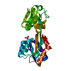
| ||||||||||||
|---|---|---|---|---|---|---|---|---|---|---|---|---|---|
| 1 |
| ||||||||||||
| Unit cell |
|
-Components
| #1: Protein | Mass: 27008.760 Da / Num. of mol.: 1 Source method: isolated from a genetically manipulated source Source: (gene. exp.) Klebsiella pneumoniae subsp. pneumoniae (strain HS11286) (bacteria)Gene: KPHS_16010 / Production host: |
|---|---|
| #2: Chemical | ChemComp-MOO /   Mass: 159.938 Da / Num. of mol.: 1 / Source method: obtained synthetically / Formula: MoO4 / Feature type: SUBJECT OF INVESTIGATION Mass: 159.938 Da / Num. of mol.: 1 / Source method: obtained synthetically / Formula: MoO4 / Feature type: SUBJECT OF INVESTIGATION |
| #3: Water | ChemComp-HOH /  Mass: 18.015 Da / Num. of mol.: 275 / Source method: isolated from a natural source / Formula: H2O Mass: 18.015 Da / Num. of mol.: 275 / Source method: isolated from a natural source / Formula: H2O |
| Has ligand of interest | Y |
-Experimental details
-Experiment
| Experiment | Method: X-RAY DIFFRACTION / Number of used crystals: 1 |
|---|
- Sample preparation
| Crystal | Density Matthews: 2.37 Å3/Da / Density % sol: 48.12 % |
|---|---|
| Crystal grow | Temperature: 289 K / Method: vapor diffusion, sitting drop / pH: 3 Details: 0.1 M Citric acid, 25% w/v Polyethylene glycol 3,350, molybdate |
-Data collection
| Diffraction | Mean temperature: 100 K / Serial crystal experiment: N |
|---|---|
| Diffraction source | Source: SYNCHROTRON / Site: SSRF / Beamline: BL19U1 / Wavelength: 0.987 Å |
| Detector | Type: DECTRIS PILATUS 6M / Detector: PIXEL / Date: May 9, 2023 |
| Radiation | Protocol: SINGLE WAVELENGTH / Monochromatic (M) / Laue (L): M / Scattering type: x-ray |
| Radiation wavelength | Wavelength: 0.987 Å / Relative weight: 1 |
| Reflection | Resolution: 1.77→50 Å / Num. obs: 20631 / % possible obs: 98.4 % / Redundancy: 6.8 % / Biso Wilson estimate: 18.64 Å2 / Rpim(I) all: 0.036 / Net I/σ(I): 16.7 |
| Reflection shell | Resolution: 1.77→1.83 Å / Num. unique obs: 2045 / Rpim(I) all: 0.149 |
- Processing
| Software |
| |||||||||||||||||||||||||||||||||||||||||||||||||||||||||||||||||||||||||||||||||||||||||||||||||||||||||
|---|---|---|---|---|---|---|---|---|---|---|---|---|---|---|---|---|---|---|---|---|---|---|---|---|---|---|---|---|---|---|---|---|---|---|---|---|---|---|---|---|---|---|---|---|---|---|---|---|---|---|---|---|---|---|---|---|---|---|---|---|---|---|---|---|---|---|---|---|---|---|---|---|---|---|---|---|---|---|---|---|---|---|---|---|---|---|---|---|---|---|---|---|---|---|---|---|---|---|---|---|---|---|---|---|---|---|
| Refinement | Method to determine structure: MOLECULAR REPLACEMENT / Resolution: 1.77→28.76 Å / SU ML: 0.1844 / Cross valid method: FREE R-VALUE / σ(F): 0.02 / Phase error: 19.7607 Stereochemistry target values: GeoStd + Monomer Library + CDL v1.2
| |||||||||||||||||||||||||||||||||||||||||||||||||||||||||||||||||||||||||||||||||||||||||||||||||||||||||
| Solvent computation | Shrinkage radii: 0.9 Å / VDW probe radii: 1.1 Å / Solvent model: FLAT BULK SOLVENT MODEL | |||||||||||||||||||||||||||||||||||||||||||||||||||||||||||||||||||||||||||||||||||||||||||||||||||||||||
| Displacement parameters | Biso mean: 21.77 Å2 | |||||||||||||||||||||||||||||||||||||||||||||||||||||||||||||||||||||||||||||||||||||||||||||||||||||||||
| Refinement step | Cycle: LAST / Resolution: 1.77→28.76 Å
| |||||||||||||||||||||||||||||||||||||||||||||||||||||||||||||||||||||||||||||||||||||||||||||||||||||||||
| Refine LS restraints |
| |||||||||||||||||||||||||||||||||||||||||||||||||||||||||||||||||||||||||||||||||||||||||||||||||||||||||
| LS refinement shell |
|
