 Movie
Movie Controller


+ Open data

- Basic information
| Entry | Database: PDB / ID: 8k84 | ||||||
|---|---|---|---|---|---|---|---|
| Title | De novo design protein -N3 | ||||||
 Components Components | De novo design protein | ||||||
 Keywords Keywords | DE NOVO PROTEIN / De novo design protein | ||||||
| Function / homology | DI(HYDROXYETHYL)ETHER Function and homology information Function and homology information | ||||||
| Biological species | synthetic construct (others) | ||||||
| Method |  X-RAY DIFFRACTION / SYNCHROTRON / MOLECULAR REPLACEMENT / Resolution: 2 Å X-RAY DIFFRACTION / SYNCHROTRON / MOLECULAR REPLACEMENT / Resolution: 2 Å | ||||||
 Authors Authors | Wang, S. / Liu, Y. | ||||||
| Funding support | 1items
| ||||||
 Citation Citation | Journal: Nat.Methods / Year: 2024 Title: De novo protein design with a denoising diffusion network independent of pretrained structure prediction models. Authors: Liu, Y. / Wang, S. / Dong, J. / Chen, L. / Wang, X. / Wang, L. / Li, F. / Wang, C. / Zhang, J. / Wang, Y. / Wei, S. / Chen, Q. / Liu, H. | ||||||
| History |
|
- Structure visualization
| Structure viewer | Molecule: MolmilJmol/JSmol |
|---|
- Downloads & links
-Download
| PDBx/mmCIF format | 8k84.cif.gz | 99.5 KB | Display | PDBx/mmCIF format |
|---|---|---|---|---|
| PDB format | pdb8k84.ent.gz | 60.9 KB | Display | PDB format |
| PDBx/mmJSON format | 8k84.json.gz | Tree view | PDBx/mmJSON format | |
| Others |  Other downloads Other downloads |
-Validation report
| Arichive directory | https://data.pdbj.org/pub/pdb/validation_reports/k8/8k84ftp://data.pdbj.org/pub/pdb/validation_reports/k8/8k84 | HTTPS FTP |
|---|
-Related structure data
| Related structure data |  8k7mC  8k7oC  8k7zC  8k83C  8k8fC  8k8gC  8k8iC  8ka6C  8ka7C  8kacC  8kc0C  8kc1C  8kc4C  8kc5C  8kc8C  8kcjC  8kckC  8kdqC  8w97C 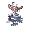 8wwcC  8wx8C C: citing same article ( |
|---|---|
| Similar structure data |
-Links
 PDBj
PDBj
- Assembly
| Deposited unit | 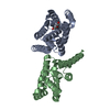
| ||||||||||||
|---|---|---|---|---|---|---|---|---|---|---|---|---|---|
| 1 | 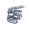
| ||||||||||||
| 2 | 
| ||||||||||||
| Unit cell |
|
-Components
| #1: Protein | Mass: 19085.541 Da / Num. of mol.: 2 Source method: isolated from a genetically manipulated source Source: (gene. exp.) synthetic construct (others) / Production host:  #2: Chemical | ChemComp-PEG / |   Mass: 106.120 Da / Num. of mol.: 1 / Source method: obtained synthetically / Formula: C4H10O3 Mass: 106.120 Da / Num. of mol.: 1 / Source method: obtained synthetically / Formula: C4H10O3#3: Water | ChemComp-HOH / |  Mass: 18.015 Da / Num. of mol.: 257 / Source method: isolated from a natural source / Formula: H2O Mass: 18.015 Da / Num. of mol.: 257 / Source method: isolated from a natural source / Formula: H2OHas ligand of interest | N | Has protein modification | N | |
|---|
-Experimental details
-Experiment
| Experiment | Method: X-RAY DIFFRACTION / Number of used crystals: 1 |
|---|
- Sample preparation
| Crystal | Density Matthews: 2.05 Å3/Da / Density % sol: 39.85 % |
|---|---|
| Crystal grow | Temperature: 289 K / Method: vapor diffusion / Details: 0.2M Imidazole malate pH5.5 33% w/v PEG600 |
-Data collection
| Diffraction | Mean temperature: 100 K / Serial crystal experiment: N |
|---|---|
| Diffraction source | Source: SYNCHROTRON / Site: SSRF  / Beamline: BL19U1 / Wavelength: 0.9785 Å / Beamline: BL19U1 / Wavelength: 0.9785 Å |
| Detector | Type: DECTRIS PILATUS3 6M / Detector: PIXEL / Date: May 13, 2023 |
| Radiation | Protocol: SINGLE WAVELENGTH / Monochromatic (M) / Laue (L): M / Scattering type: x-ray |
| Radiation wavelength | Wavelength: 0.9785 Å / Relative weight: 1 |
| Reflection | Resolution: 2→59.82 Å / Num. obs: 21086 / % possible obs: 99.3 % / Redundancy: 3 % / Biso Wilson estimate: 19.05 Å2 / CC1/2: 0.99 / Net I/σ(I): 6.7 |
| Reflection shell | Resolution: 2→2.05 Å / Num. unique obs: 1527 / CC1/2: 0.584 |
- Processing
| Software |
| |||||||||||||||||||||||||||||||||||||||||||||||||||||||||||||||
|---|---|---|---|---|---|---|---|---|---|---|---|---|---|---|---|---|---|---|---|---|---|---|---|---|---|---|---|---|---|---|---|---|---|---|---|---|---|---|---|---|---|---|---|---|---|---|---|---|---|---|---|---|---|---|---|---|---|---|---|---|---|---|---|---|
| Refinement | Method to determine structure: MOLECULAR REPLACEMENT / Resolution: 2→25.01 Å / SU ML: 0.2577 / Cross valid method: FREE R-VALUE / σ(F): 1.34 / Phase error: 24.7565 Stereochemistry target values: GeoStd + Monomer Library + CDL v1.2
| |||||||||||||||||||||||||||||||||||||||||||||||||||||||||||||||
| Solvent computation | Shrinkage radii: 0.9 Å / VDW probe radii: 1.1 Å / Solvent model: FLAT BULK SOLVENT MODEL | |||||||||||||||||||||||||||||||||||||||||||||||||||||||||||||||
| Displacement parameters | Biso mean: 20.91 Å2 | |||||||||||||||||||||||||||||||||||||||||||||||||||||||||||||||
| Refinement step | Cycle: LAST / Resolution: 2→25.01 Å
| |||||||||||||||||||||||||||||||||||||||||||||||||||||||||||||||
| Refine LS restraints |
| |||||||||||||||||||||||||||||||||||||||||||||||||||||||||||||||
| LS refinement shell |
|
