 Movie
Movie Controller

[English] 日本語
 Yorodumi
- PDB-8k6u: Serial Femtosecond X-ray structure of E.coli Cyanase with un-mode... -
+ Open data

- Basic information
| Entry | Database: PDB / ID: 8k6u | ||||||
|---|---|---|---|---|---|---|---|
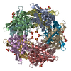
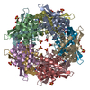
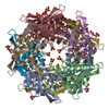
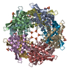
| Title | Serial Femtosecond X-ray structure of E.coli Cyanase with un-modeled density at active site | ||||||
 Components Components | Cyanate hydratase | ||||||
 Keywords Keywords | LYASE / bi-substrate enzyme / enzyme reaction / room temperature | ||||||
| Function / homology |  Function and homology information Function and homology informationcyanate catabolic process / cyanase / cyanate hydratase activity / DNA binding Similarity search - Function | ||||||
| Biological species |  | ||||||
| Method |  X-RAY DIFFRACTION / FREE ELECTRON LASER / MOLECULAR REPLACEMENT / Resolution: 1.9 Å X-RAY DIFFRACTION / FREE ELECTRON LASER / MOLECULAR REPLACEMENT / Resolution: 1.9 Å | ||||||
 Authors Authors | Kim, J. / Nam, K.H. / Cho, Y. | ||||||
| Funding support |  Korea, Republic Of, 1items Korea, Republic Of, 1items
| ||||||
 Citation Citation | Journal: Acta Crystallogr D Struct Biol / Year: 2023 Title: Structural mechanism of Escherichia coli cyanase. Authors: Kim, J. / Kim, Y. / Park, J. / Nam, K.H. / Cho, Y. | ||||||
| History |
|
- Structure visualization
| Structure viewer | Molecule: MolmilJmol/JSmol |
|---|
- Downloads & links
-Download
| PDBx/mmCIF format | 8k6u.cif.gz | 311.3 KB | Display | PDBx/mmCIF format |
|---|---|---|---|---|
| PDB format | pdb8k6u.ent.gz | 252.9 KB | Display | PDB format |
| PDBx/mmJSON format | 8k6u.json.gz | Tree view | PDBx/mmJSON format | |
| Others |  Other downloads Other downloads |
-Validation report
| Arichive directory | https://data.pdbj.org/pub/pdb/validation_reports/k6/8k6uftp://data.pdbj.org/pub/pdb/validation_reports/k6/8k6u | HTTPS FTP |
|---|
-Related structure data
-Links
 PDBj
PDBj


- Assembly
| Deposited unit | 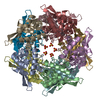
| ||||||||
|---|---|---|---|---|---|---|---|---|---|
| 1 |
| ||||||||
| Unit cell |
|
-Components
| #1: Protein | Mass: 17481.266 Da / Num. of mol.: 10 Source method: isolated from a genetically manipulated source Source: (gene. exp.) #2: Chemical | ChemComp-SO4 /   Mass: 96.063 Da / Num. of mol.: 5 / Source method: obtained synthetically / Formula: SO4 Mass: 96.063 Da / Num. of mol.: 5 / Source method: obtained synthetically / Formula: SO4#3: Water | ChemComp-HOH / |  Mass: 18.015 Da / Num. of mol.: 603 / Source method: isolated from a natural source / Formula: H2O Mass: 18.015 Da / Num. of mol.: 603 / Source method: isolated from a natural source / Formula: H2OHas ligand of interest | N | |
|---|
-Experimental details
-Experiment
| Experiment | Method: X-RAY DIFFRACTION / Number of used crystals: 1 |
|---|
- Sample preparation
| Crystal | Density Matthews: 2.56 Å3/Da / Density % sol: 51.87 % |
|---|---|
| Crystal grow | Temperature: 293 K / Method: batch mode / pH: 7.3 Details: 50mM Tris-Cl (pH 7.3), 50 mM potassium phosphate, and 2.5 M ammonium sulfate |
-Data collection
| Diffraction | Mean temperature: 293 K / Serial crystal experiment: Y |
|---|---|
| Diffraction source | Source: FREE ELECTRON LASER / Site: PAL-XFEL / Beamline: NCI / Wavelength: 1.305 Å |
| Detector | Type: RAYONIX MX225-HS / Detector: CCD / Date: May 22, 2022 |
| Radiation | Protocol: SINGLE WAVELENGTH / Monochromatic (M) / Laue (L): M / Scattering type: x-ray |
| Radiation wavelength | Wavelength: 1.305 Å / Relative weight: 1 |
| Reflection | Resolution: 1.9→75.9 Å / Num. obs: 266307 / % possible obs: 100 % / Redundancy: 295.4 % / CC1/2: 0.96 / CC star: 0.99 / R split: 0.18 / Net I/σ(I): 4.54 |
| Reflection shell | Resolution: 1.9→2 Å / Mean I/σ(I) obs: 1.41 / Num. unique obs: 18842 / CC1/2: 0.35 / R split: 0.83 |
- Processing
| Software |
| |||||||||||||||||||||||||||||||||||||||||||||||||||||||||||||||||||||||||||||||||||||||||||||||||||||||||
|---|---|---|---|---|---|---|---|---|---|---|---|---|---|---|---|---|---|---|---|---|---|---|---|---|---|---|---|---|---|---|---|---|---|---|---|---|---|---|---|---|---|---|---|---|---|---|---|---|---|---|---|---|---|---|---|---|---|---|---|---|---|---|---|---|---|---|---|---|---|---|---|---|---|---|---|---|---|---|---|---|---|---|---|---|---|---|---|---|---|---|---|---|---|---|---|---|---|---|---|---|---|---|---|---|---|---|
| Refinement | Method to determine structure: MOLECULAR REPLACEMENT / Resolution: 1.9→57.13 Å / SU ML: 0.23 / Cross valid method: FREE R-VALUE / σ(F): 1.96 / Phase error: 22.21 / Stereochemistry target values: ML
| |||||||||||||||||||||||||||||||||||||||||||||||||||||||||||||||||||||||||||||||||||||||||||||||||||||||||
| Solvent computation | Shrinkage radii: 0.9 Å / VDW probe radii: 1.11 Å / Solvent model: FLAT BULK SOLVENT MODEL | |||||||||||||||||||||||||||||||||||||||||||||||||||||||||||||||||||||||||||||||||||||||||||||||||||||||||
| Refinement step | Cycle: LAST / Resolution: 1.9→57.13 Å
| |||||||||||||||||||||||||||||||||||||||||||||||||||||||||||||||||||||||||||||||||||||||||||||||||||||||||
| Refine LS restraints |
| |||||||||||||||||||||||||||||||||||||||||||||||||||||||||||||||||||||||||||||||||||||||||||||||||||||||||
| LS refinement shell |
|
