 Movie
Movie Controller

[English] 日本語
 Yorodumi
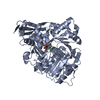
- PDB-8k5s: The structure of EntE with 3-(prop-2-yn-1-yloxy)benzoic acid sulf... -
+ Open data

- Basic information
| Entry | Database: PDB / ID: 8k5s | ||||||
|---|---|---|---|---|---|---|---|
| Title | The structure of EntE with 3-(prop-2-yn-1-yloxy)benzoic acid sulfamoyl adenosine | ||||||
 Components Components | Enterobactin synthase component E | ||||||
 Keywords Keywords | LIGASE / Adenylation / ATP binding / Nonribosomal peptide synthetase / Biosynthesis | ||||||
| Function / homology |  Function and homology information Function and homology information2,3-dihydroxybenzoate-[aryl-carrier protein] ligase / enterobactin synthase / 2,3-dihydroxybenzoate--[aryl-carrier protein] ligase / 2,3-dihydroxybenzoate-serine ligase activity / enterobactin biosynthetic process / acyltransferase activity / ATP binding / membrane / cytosol Similarity search - Function | ||||||
| Biological species |  | ||||||
| Method |  X-RAY DIFFRACTION / SYNCHROTRON / MOLECULAR REPLACEMENT / Resolution: 2.65 Å X-RAY DIFFRACTION / SYNCHROTRON / MOLECULAR REPLACEMENT / Resolution: 2.65 Å | ||||||
 Authors Authors | Miyanaga, A. / Ishikawa, F. | ||||||
| Funding support | 1items
| ||||||
 Citation Citation | Journal: Acs Chem.Biol. / Year: 2024 Title: Biosynthetic Incorporation of Non-native Aryl Acid Building Blocks into Peptide Products Using Engineered Adenylation Domains. Authors: Ishikawa, F. / Nohara, M. / Miyanaga, A. / Kuramoto, S. / Miyano, N. / Asamizu, S. / Kudo, F. / Onaka, H. / Eguchi, T. / Tanabe, G. | ||||||
| History |
|
- Structure visualization
| Structure viewer | Molecule: MolmilJmol/JSmol |
|---|
- Downloads & links
-Download
| PDBx/mmCIF format | 8k5s.cif.gz | 212.3 KB | Display | PDBx/mmCIF format |
|---|---|---|---|---|
| PDB format | pdb8k5s.ent.gz | 169.1 KB | Display | PDB format |
| PDBx/mmJSON format | 8k5s.json.gz | Tree view | PDBx/mmJSON format | |
| Others |  Other downloads Other downloads |
-Validation report
| Arichive directory | https://data.pdbj.org/pub/pdb/validation_reports/k5/8k5sftp://data.pdbj.org/pub/pdb/validation_reports/k5/8k5s | HTTPS FTP |
|---|
-Related structure data
-Links
 PDBj
PDBj
- Assembly
| Deposited unit | 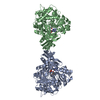
| ||||||||||||||||||
|---|---|---|---|---|---|---|---|---|---|---|---|---|---|---|---|---|---|---|---|
| 1 |
| ||||||||||||||||||
| Unit cell |
| ||||||||||||||||||
| Noncrystallographic symmetry (NCS) | NCS domain:
NCS domain segments: Component-ID: _ / Ens-ID: 1 / Beg auth comp-ID: ILE / Beg label comp-ID: ILE / End auth comp-ID: GLN / End label comp-ID: GLN / Refine code: _ / Auth seq-ID: 3 - 528 / Label seq-ID: 3 - 528
|
-Components
| #1: Protein | Mass: 61333.227 Da / Num. of mol.: 2 / Mutation: N235G Source method: isolated from a genetically manipulated source Source: (gene. exp.) References: UniProt: P10378, enterobactin synthase, 2,3-dihydroxybenzoate-[aryl-carrier protein] ligase #2: Chemical | Mass: 504.473 Da / Num. of mol.: 2 / Source method: obtained synthetically / Formula: C20H20N6O8S / Feature type: SUBJECT OF INVESTIGATION #3: Water | ChemComp-HOH / |  Mass: 18.015 Da / Num. of mol.: 62 / Source method: isolated from a natural source / Formula: H2O Mass: 18.015 Da / Num. of mol.: 62 / Source method: isolated from a natural source / Formula: H2OHas ligand of interest | Y | Has protein modification | N | |
|---|
-Experimental details
-Experiment
| Experiment | Method: X-RAY DIFFRACTION / Number of used crystals: 1 |
|---|
- Sample preparation
| Crystal | Density Matthews: 2.48 Å3/Da / Density % sol: 50.43 % |
|---|---|
| Crystal grow | Temperature: 293 K / Method: vapor diffusion, sitting drop / pH: 7.5 / Details: HEPES-Na, MPD |
-Data collection
| Diffraction | Mean temperature: 100 K / Serial crystal experiment: N |
|---|---|
| Diffraction source | Source: SYNCHROTRON / Site: Photon Factory  / Beamline: AR-NW12A / Wavelength: 1 Å / Beamline: AR-NW12A / Wavelength: 1 Å |
| Detector | Type: DECTRIS PILATUS3 S 2M / Detector: PIXEL / Date: Nov 7, 2020 |
| Radiation | Monochromator: Numerical link type Si(111) double crystal monochromator Protocol: SINGLE WAVELENGTH / Monochromatic (M) / Laue (L): M / Scattering type: x-ray |
| Radiation wavelength | Wavelength: 1 Å / Relative weight: 1 |
| Reflection | Resolution: 2.65→50 Å / Num. obs: 35229 / % possible obs: 99.5 % / Redundancy: 3.4 % / CC1/2: 0.993 / Rmerge(I) obs: 0.102 / Net I/σ(I): 5.9 |
| Reflection shell | Resolution: 2.65→2.8 Å / Redundancy: 3.5 % / Rmerge(I) obs: 0.468 / Mean I/σ(I) obs: 1.9 / Num. unique obs: 4630 / CC1/2: 0.832 / % possible all: 99.1 |
- Processing
| Software |
| ||||||||||||||||||||||||||||||||||||||||||||||||||||||||||||
|---|---|---|---|---|---|---|---|---|---|---|---|---|---|---|---|---|---|---|---|---|---|---|---|---|---|---|---|---|---|---|---|---|---|---|---|---|---|---|---|---|---|---|---|---|---|---|---|---|---|---|---|---|---|---|---|---|---|---|---|---|---|
| Refinement | Method to determine structure: MOLECULAR REPLACEMENT / Resolution: 2.65→45.56 Å / Cor.coef. Fo:Fc: 0.948 / Cor.coef. Fo:Fc free: 0.927 / SU B: 11.971 / SU ML: 0.243 / Cross valid method: THROUGHOUT / σ(F): 0 / ESU R: 2.289 / ESU R Free: 0.294 / Stereochemistry target values: MAXIMUM LIKELIHOOD Details: HYDROGENS HAVE BEEN ADDED IN THE RIDING POSITIONS U VALUES : REFINED INDIVIDUALLY
| ||||||||||||||||||||||||||||||||||||||||||||||||||||||||||||
| Solvent computation | Ion probe radii: 0.8 Å / Shrinkage radii: 0.8 Å / VDW probe radii: 1.2 Å / Solvent model: MASK | ||||||||||||||||||||||||||||||||||||||||||||||||||||||||||||
| Displacement parameters | Biso max: 133.61 Å2 / Biso mean: 35.236 Å2 / Biso min: 8.4 Å2
| ||||||||||||||||||||||||||||||||||||||||||||||||||||||||||||
| Refinement step | Cycle: final / Resolution: 2.65→45.56 Å
| ||||||||||||||||||||||||||||||||||||||||||||||||||||||||||||
| Refine LS restraints |
| ||||||||||||||||||||||||||||||||||||||||||||||||||||||||||||
| Refine LS restraints NCS | Ens-ID: 1 / Number: 16694 / Refine-ID: X-RAY DIFFRACTION / Type: interatomic distance / Rms dev position: 0.06 Å / Weight position: 0.05
| ||||||||||||||||||||||||||||||||||||||||||||||||||||||||||||
| LS refinement shell | Resolution: 2.65→2.719 Å / Rfactor Rfree error: 0 / Total num. of bins used: 20
|
