 Movie
Movie Controller


+ Open data

- Basic information
| Entry | Database: PDB / ID: 8k35 | |||||||||||||||||||||||||||||||||
|---|---|---|---|---|---|---|---|---|---|---|---|---|---|---|---|---|---|---|---|---|---|---|---|---|---|---|---|---|---|---|---|---|---|---|
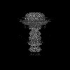
| Title | Structure of the bacteriophage lambda tail tip complex | |||||||||||||||||||||||||||||||||
 Components Components |
| |||||||||||||||||||||||||||||||||
 Keywords Keywords | VIRAL PROTEIN / Complex | |||||||||||||||||||||||||||||||||
| Function / homology |  Function and homology information Function and homology informationvirus tail, tube / symbiont genome ejection through host cell envelope, long flexible tail mechanism / viral tail assembly / virus tail / 4 iron, 4 sulfur cluster binding / host cell cytoplasm / entry receptor-mediated virion attachment to host cell / receptor-mediated virion attachment to host cell / virion attachment to host cell / metal ion binding Similarity search - Function | |||||||||||||||||||||||||||||||||
| Biological species |  Escherichia phage Lambda (virus) Escherichia phage Lambda (virus) | |||||||||||||||||||||||||||||||||
| Method | ELECTRON MICROSCOPY / single particle reconstruction / cryo EM / Resolution: 3.44 Å | |||||||||||||||||||||||||||||||||
 Authors Authors | Xiao, H. / Tan, L. / Cheng, L.P. / Liu, H.R. | |||||||||||||||||||||||||||||||||
| Funding support |  China, 4items China, 4items
| |||||||||||||||||||||||||||||||||
 Citation Citation | Journal: PLoS Biol / Year: 2023 Title: Structure of the siphophage neck-Tail complex suggests that conserved tail tip proteins facilitate receptor binding and tail assembly. Authors: Hao Xiao / Le Tan / Zhixue Tan / Yewei Zhang / Wenyuan Chen / Xiaowu Li / Jingdong Song / Lingpeng Cheng / Hongrong Liu / Abstract: Siphophages have a long, flexible, and noncontractile tail that connects to the capsid through a neck. The phage tail is essential for host cell recognition and virus-host cell interactions; ...Siphophages have a long, flexible, and noncontractile tail that connects to the capsid through a neck. The phage tail is essential for host cell recognition and virus-host cell interactions; moreover, it serves as a channel for genome delivery during infection. However, the in situ high-resolution structure of the neck-tail complex of siphophages remains unknown. Here, we present the structure of the siphophage lambda "wild type," the most widely used, laboratory-adapted fiberless mutant. The neck-tail complex comprises a channel formed by stacked 12-fold and hexameric rings and a 3-fold symmetrical tip. The interactions among DNA and a total of 246 tail protein molecules forming the tail and neck have been characterized. Structural comparisons of the tail tips, the most diversified region across the lambda and other long-tailed phages or tail-like machines, suggest that their tail tip contains conserved domains, which facilitate tail assembly, receptor binding, cell adsorption, and DNA retaining/releasing. These domains are distributed in different tail tip proteins in different phages or tail-like machines. The side tail fibers are not required for the phage particle to orient itself vertically to the surface of the host cell during attachment. | |||||||||||||||||||||||||||||||||
| History |
|
- Structure visualization
| Structure viewer | Molecule: MolmilJmol/JSmol |
|---|
- Downloads & links
-Download
| PDBx/mmCIF format | 8k35.cif.gz | 939 KB | Display | PDBx/mmCIF format |
|---|---|---|---|---|
| PDB format | pdb8k35.ent.gz | Display | PDB format | |
| PDBx/mmJSON format | 8k35.json.gz | Tree view | PDBx/mmJSON format | |
| Others |  Other downloads Other downloads |
-Validation report
| Arichive directory | https://data.pdbj.org/pub/pdb/validation_reports/k3/8k35ftp://data.pdbj.org/pub/pdb/validation_reports/k3/8k35 | HTTPS FTP |
|---|
-Related structure data
| Related structure data | 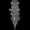 36844MC 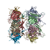 8k36C  8k37C  8k38C  8k39C M: map data used to model this data C: citing same article ( |
|---|---|
| Similar structure data |

-Links
 PDBj
PDBj


- Assembly
| Deposited unit | 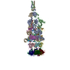
|
|---|---|
| 1 |
|
-Components
-Protein , 4 types, 15 molecules IABxKQLRTUVWPMS
| #1: Protein | Mass: 124550.625 Da / Num. of mol.: 3 / Source method: isolated from a natural source / Source: (natural) Escherichia phage Lambda (virus) / References: UniProt: P03749#4: Protein | Mass: 23146.590 Da / Num. of mol.: 3 / Source method: isolated from a natural source / Source: (natural) Escherichia phage Lambda (virus) / References: UniProt: P03730#5: Protein | Mass: 25831.779 Da / Num. of mol.: 6 / Source method: isolated from a natural source / Source: (natural) Escherichia phage Lambda (virus) / References: UniProt: P03733#6: Protein | Mass: 92393.430 Da / Num. of mol.: 3 / Source method: isolated from a natural source / Source: (natural) Escherichia phage Lambda (virus) / References: UniProt: P03736 |
|---|
-Tail tip protein ... , 2 types, 9 molecules JGNHOCEDF
| #2: Protein | Mass: 25730.578 Da / Num. of mol.: 3 / Source method: isolated from a natural source / Source: (natural) Escherichia phage Lambda (virus) / References: UniProt: P03738#3: Protein | Mass: 12547.373 Da / Num. of mol.: 6 / Source method: isolated from a natural source / Source: (natural) Escherichia phage Lambda (virus) / References: UniProt: P03737 |
|---|
-Non-polymers , 1 types, 3 molecules 
| #7: Chemical |  Mass: 351.640 Da / Num. of mol.: 3 / Source method: obtained synthetically / Formula: Fe4S4 Mass: 351.640 Da / Num. of mol.: 3 / Source method: obtained synthetically / Formula: Fe4S4 |
|---|
-Details
| Has ligand of interest | N |
|---|---|
| Has protein modification | N |
-Experimental details
-Experiment
| Experiment | Method: ELECTRON MICROSCOPY |
|---|---|
| EM experiment | Aggregation state: PARTICLE / 3D reconstruction method: single particle reconstruction |
- Sample preparation
| Component | Name: Escherichia phage Lambda / Type: VIRUS / Entity ID: #1-#6 / Source: NATURAL |
|---|---|
| Source (natural) | Organism: Escherichia phage Lambda (virus) |
| Details of virus | Empty: NO / Enveloped: NO / Isolate: SPECIES / Type: VIRION |
| Buffer solution | pH: 7.4 |
| Specimen | Embedding applied: NO / Shadowing applied: NO / Staining applied: NO / Vitrification applied: YES |
| Vitrification | Cryogen name: ETHANE |
- Electron microscopy imaging
| Experimental equipment |  Model: Titan Krios / Image courtesy: FEI Company |
|---|---|
| Microscopy | Model: FEI TITAN KRIOS |
| Electron gun | Electron source:  FIELD EMISSION GUN / Accelerating voltage: 300 kV / Illumination mode: FLOOD BEAM FIELD EMISSION GUN / Accelerating voltage: 300 kV / Illumination mode: FLOOD BEAM |
| Electron lens | Mode: BRIGHT FIELD / Nominal defocus max: 2200 nm / Nominal defocus min: 1600 nm |
| Image recording | Electron dose: 32 e/Å2 / Film or detector model: GATAN K3 (6k x 4k) |
- Processing
| EM software | Name: PHENIX / Category: model refinement | ||||||||||||||||||||||||
|---|---|---|---|---|---|---|---|---|---|---|---|---|---|---|---|---|---|---|---|---|---|---|---|---|---|
| CTF correction | Type: PHASE FLIPPING ONLY | ||||||||||||||||||||||||
| 3D reconstruction | Resolution: 3.44 Å / Resolution method: FSC 0.143 CUT-OFF / Num. of particles: 54385 / Symmetry type: POINT | ||||||||||||||||||||||||
| Refine LS restraints |
|
