 Movie
Movie Controller


+ Open data

- Basic information
| Entry | Database: PDB / ID: 8jzw | ||||||
|---|---|---|---|---|---|---|---|
| Title | Cystal structure of HP1 in complex with TRIM66 peptide | ||||||
 Components Components |
| ||||||
 Keywords Keywords | PROTEIN BINDING / TRIM66 / HP1 | ||||||
| Function / homology |  Function and homology information Function and homology information: / pericentric heterochromatin / chromatin organization / chromatin binding / negative regulation of transcription by RNA polymerase II / nucleus Similarity search - Function | ||||||
| Biological species |  Homo sapiens (human) Homo sapiens (human) | ||||||
| Method |  X-RAY DIFFRACTION / SYNCHROTRON / MOLECULAR REPLACEMENT / Resolution: 1.8 Å X-RAY DIFFRACTION / SYNCHROTRON / MOLECULAR REPLACEMENT / Resolution: 1.8 Å | ||||||
 Authors Authors | Shen, S.Y. / Li, F.D. / Shi, Y.Y. | ||||||
| Funding support |  China, 1items China, 1items
| ||||||
 Citation Citation | Journal: To Be Published Title: Cystal structure of HP1 in complex with TRIM66 peptide Authors: Shen, S.Y. / Li, F.D. / Shi, Y.Y. | ||||||
| History |
|
- Structure visualization
| Structure viewer | Molecule: MolmilJmol/JSmol |
|---|
- Downloads & links
-Download
| PDBx/mmCIF format | 8jzw.cif.gz | 128 KB | Display | PDBx/mmCIF format |
|---|---|---|---|---|
| PDB format | pdb8jzw.ent.gz | 99.6 KB | Display | PDB format |
| PDBx/mmJSON format | 8jzw.json.gz | Tree view | PDBx/mmJSON format | |
| Others |  Other downloads Other downloads |
-Validation report
| Summary document | 8jzw_validation.pdf.gz | 453 KB | Display | wwPDB validaton report |
|---|---|---|---|---|
| Full document | 8jzw_full_validation.pdf.gz | 453.9 KB | Display | |
| Data in XML | 8jzw_validation.xml.gz | 13.8 KB | Display | |
| Data in CIF | 8jzw_validation.cif.gz | 20.2 KB | Display | |
| Arichive directory | https://data.pdbj.org/pub/pdb/validation_reports/jz/8jzwftp://data.pdbj.org/pub/pdb/validation_reports/jz/8jzw | HTTPS FTP |
-Related structure data
| Similar structure data |
|---|
-Links
 PDBj
PDBj- Assembly
| Deposited unit | 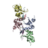
| ||||||||
|---|---|---|---|---|---|---|---|---|---|
| 1 | 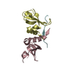
| ||||||||
| 2 | 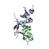
| ||||||||
| Unit cell |
|
-Components
| #1: Protein | Mass: 9185.379 Da / Num. of mol.: 4 Source method: isolated from a genetically manipulated source Source: (gene. exp.) Homo sapiens (human) / Gene: CBX3, CCDC32, hCG_1745364, hCG_1999239, tcag7.173 / Production host:  #2: Protein/peptide | Mass: 2174.589 Da / Num. of mol.: 2 / Source method: obtained synthetically / Source: (synth.) Homo sapiens (human)#3: Water | ChemComp-HOH / |  Mass: 18.015 Da / Num. of mol.: 225 / Source method: isolated from a natural source / Formula: H2O Mass: 18.015 Da / Num. of mol.: 225 / Source method: isolated from a natural source / Formula: H2O |
|---|
-Experimental details
-Experiment
| Experiment | Method: X-RAY DIFFRACTION / Number of used crystals: 1 |
|---|
- Sample preparation
| Crystal | Density Matthews: 1.76 Å3/Da / Density % sol: 30.05 % |
|---|---|
| Crystal grow | Temperature: 293.15 K / Method: batch mode Details: 30% PEG MME 2000, 0.1 mmol/L sodium cacodylate (pH 6.0) |
-Data collection
| Diffraction | Mean temperature: 293.15 K / Serial crystal experiment: N |
|---|---|
| Diffraction source | Source: SYNCHROTRON / Site: SSRF / Beamline: BL19U1 / Wavelength: 0.979 Å |
| Detector | Type: DECTRIS PILATUS3 6M / Detector: PIXEL / Date: Apr 20, 2017 |
| Diffraction measurement | Details: 1.00 degrees, 0.1 sec, detector distance 200.00 mm / Method: \w scans |
| Radiation | Protocol: MAD / Monochromatic (M) / Laue (L): M / Scattering type: x-ray |
| Radiation wavelength | Wavelength: 0.979 Å / Relative weight: 1 |
| Reflection | Av R equivalents: 0.091 / Number: 176397 |
| Reflection | Resolution: 1.8→40 Å / Num. obs: 25428 / % possible obs: 96.5 % / Observed criterion σ(F): 0 / Observed criterion σ(I): -3 / Redundancy: 6.9 % / Rmerge(I) obs: 0.091 / Rsym value: 0.091 / Net I/av σ(I): 15.545 / Net I/σ(I): 6.9 |
| Reflection shell | Resolution: 1.8→1.86 Å / Redundancy: 6.8 % / Rmerge(I) obs: 0.576 / Mean I/σ(I) obs: 2.188 / Num. unique obs: 2491 / Rsym value: 0.576 / % possible all: 94.9 |
| Cell measurement | Reflection used: 176397 |
- Processing
| Software |
| ||||||||||||||||||||||||||||||||||||||||||||||||||||||||||||||||||||||
|---|---|---|---|---|---|---|---|---|---|---|---|---|---|---|---|---|---|---|---|---|---|---|---|---|---|---|---|---|---|---|---|---|---|---|---|---|---|---|---|---|---|---|---|---|---|---|---|---|---|---|---|---|---|---|---|---|---|---|---|---|---|---|---|---|---|---|---|---|---|---|---|
| Refinement | Method to determine structure: MOLECULAR REPLACEMENT / Resolution: 1.8→35.94 Å / SU ML: 0.18 / Cross valid method: FREE R-VALUE / σ(F): 1.35 / Phase error: 22.37 / Stereochemistry target values: ML
| ||||||||||||||||||||||||||||||||||||||||||||||||||||||||||||||||||||||
| Solvent computation | Shrinkage radii: 0.9 Å / VDW probe radii: 1.11 Å / Solvent model: FLAT BULK SOLVENT MODEL | ||||||||||||||||||||||||||||||||||||||||||||||||||||||||||||||||||||||
| Refinement step | Cycle: LAST / Resolution: 1.8→35.94 Å
| ||||||||||||||||||||||||||||||||||||||||||||||||||||||||||||||||||||||
| Refine LS restraints |
| ||||||||||||||||||||||||||||||||||||||||||||||||||||||||||||||||||||||
| LS refinement shell |
| ||||||||||||||||||||||||||||||||||||||||||||||||||||||||||||||||||||||
| Refinement TLS params. | Method: refined / Origin x: 35.4826 Å / Origin y: 25.5392 Å / Origin z: 34.1389 Å
| ||||||||||||||||||||||||||||||||||||||||||||||||||||||||||||||||||||||
| Refinement TLS group | Selection details: all |
