 Movie
Movie Controller

[English] 日本語
 Yorodumi
- PDB-8jyx: Crystal structure of the gasdermin-like protein RCD-1-1 from Neur... -
+ Open data

- Basic information
| Entry | Database: PDB / ID: 8jyx | ||||||
|---|---|---|---|---|---|---|---|
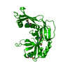
| Title | Crystal structure of the gasdermin-like protein RCD-1-1 from Neurospora crassa | ||||||
 Components Components | Maltodextrin-binding protein,Gasdermin-like protein rcd-1-1 | ||||||
 Keywords Keywords | IMMUNE SYSTEM / Pyroptosis / Gasdermnin / Allorecognition | ||||||
| Function / homology |  Function and homology information Function and homology informationwide pore channel activity / programmed cell death / carbohydrate transmembrane transporter activity / outer membrane-bounded periplasmic space / protein heterodimerization activity / plasma membrane / cytoplasm Similarity search - Function | ||||||
| Biological species |   Neurospora crassa (fungus) Neurospora crassa (fungus) | ||||||
| Method |  X-RAY DIFFRACTION / SYNCHROTRON / MOLECULAR REPLACEMENT / Resolution: 2.35 Å X-RAY DIFFRACTION / SYNCHROTRON / MOLECULAR REPLACEMENT / Resolution: 2.35 Å | ||||||
 Authors Authors | Li, Y. / Hou, Y.J. / Ding, J. | ||||||
| Funding support |  China, 1items China, 1items
| ||||||
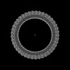
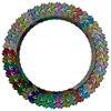
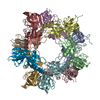
 Citation Citation | Journal: Science / Year: 2024 Title: Cleavage-independent activation of ancient eukaryotic gasdermins and structural mechanisms. Authors: Yueyue Li / Yanjie Hou / Qi Sun / Huan Zeng / Fanyi Meng / Xiang Tian / Qun He / Feng Shao / Jingjin Ding / Abstract: Gasdermins (GSDMs) are pore-forming proteins that execute pyroptosis for immune defense. GSDMs are two-domain proteins activated by proteolytic removal of the inhibitory domain. In this work, we ...Gasdermins (GSDMs) are pore-forming proteins that execute pyroptosis for immune defense. GSDMs are two-domain proteins activated by proteolytic removal of the inhibitory domain. In this work, we report two types of cleavage-independent GSDM activation. First, GSDM, a pore-forming domain-only protein from the basal metazoan , is a disulfides-linked autoinhibited dimer activated by reduction of the disulfides. The cryo-electron microscopy (cryo-EM) structure illustrates the assembly mechanism for the 44-mer GSDM pore. Second, RCD-1-1 and RCD-1-2, encoded by the polymorphic () gene in filamentous fungus , are also pore-forming domain-only GSDMs. RCD-1-1 and RCD-1-2, when encountering each other, form pores and cause pyroptosis, underlying allorecognition in . The cryo-EM structure reveals a pore of 11 RCD-1-1/RCD-1-2 heterodimers and a heterodimerization-triggered pore assembly mechanism. This study shows mechanistic diversities in GSDM activation and indicates versatile functions of GSDMs. | ||||||
| History |
|
- Structure visualization
| Structure viewer | Molecule: MolmilJmol/JSmol |
|---|
- Downloads & links
-Download
| PDBx/mmCIF format | 8jyx.cif.gz | 238.7 KB | Display | PDBx/mmCIF format |
|---|---|---|---|---|
| PDB format | pdb8jyx.ent.gz | 184.4 KB | Display | PDB format |
| PDBx/mmJSON format | 8jyx.json.gz | Tree view | PDBx/mmJSON format | |
| Others |  Other downloads Other downloads |
-Validation report
| Summary document | 8jyx_validation.pdf.gz | 1.2 MB | Display | wwPDB validaton report |
|---|---|---|---|---|
| Full document | 8jyx_full_validation.pdf.gz | 1.2 MB | Display | |
| Data in XML | 8jyx_validation.xml.gz | 39.3 KB | Display | |
| Data in CIF | 8jyx_validation.cif.gz | 54.7 KB | Display | |
| Arichive directory | https://data.pdbj.org/pub/pdb/validation_reports/jy/8jyxftp://data.pdbj.org/pub/pdb/validation_reports/jy/8jyx | HTTPS FTP |
-Related structure data


-Links
 PDBj
PDBj


- Assembly
| Deposited unit | 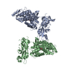
| ||||||||||||
|---|---|---|---|---|---|---|---|---|---|---|---|---|---|
| 1 | 
| ||||||||||||
| 2 | 
| ||||||||||||
| Unit cell |
|
-Components
| #1: Protein | Mass: 69615.359 Da / Num. of mol.: 2 Mutation: D108A, K109A, E198A, N199A, K265A, K388A, D389A, K174A, K175A, K176A Source method: isolated from a genetically manipulated source Source: (gene. exp.) Neurospora crassa (fungus)Gene: malE, NCTC8450_00456, NCTC9775_03059, rcd-1-1, NCU05712 Production host: #2: Polysaccharide |   Source method: isolated from a genetically manipulated source Details: oligosaccharide / References: alpha-maltose #3: Water | ChemComp-HOH / |  Mass: 18.015 Da / Num. of mol.: 162 / Source method: isolated from a natural source / Formula: H2O Mass: 18.015 Da / Num. of mol.: 162 / Source method: isolated from a natural source / Formula: H2OHas ligand of interest | N | |
|---|
-Experimental details
-Experiment
| Experiment | Method: X-RAY DIFFRACTION / Number of used crystals: 1 |
|---|
- Sample preparation
| Crystal | Density Matthews: 2.78 Å3/Da / Density % sol: 55.84 % |
|---|---|
| Crystal grow | Temperature: 291 K / Method: vapor diffusion, sitting drop / pH: 7.4 Details: 0.1 M Bicine pH 7.4, 11% PEG 3350, 3% (w/v) D(+)-Glucose monohydrate |
-Data collection
| Diffraction | Mean temperature: 100 K / Serial crystal experiment: N |
|---|---|
| Diffraction source | Source: SYNCHROTRON / Site: SSRF / Beamline: BL18U1 / Wavelength: 0.97915 Å |
| Detector | Type: DECTRIS PILATUS3 6M / Detector: PIXEL / Date: Nov 25, 2022 |
| Radiation | Protocol: SINGLE WAVELENGTH / Monochromatic (M) / Laue (L): M / Scattering type: x-ray |
| Radiation wavelength | Wavelength: 0.97915 Å / Relative weight: 1 |
| Reflection | Resolution: 2.35→79.55 Å / Num. obs: 63588 / % possible obs: 99.9 % / Redundancy: 6.8 % / Biso Wilson estimate: 45.4 Å2 / CC1/2: 0.998 / Rmerge(I) obs: 0.09 / Rpim(I) all: 0.037 / Rrim(I) all: 0.098 / Net I/σ(I): 12.3 |
| Reflection shell | Resolution: 2.35→2.41 Å / Redundancy: 6.9 % / Rmerge(I) obs: 0.82 / Mean I/σ(I) obs: 2.2 / Num. unique obs: 4459 / CC1/2: 0.881 / Rpim(I) all: 0.465 / Rrim(I) all: 0.886 / % possible all: 100 |
- Processing
| Software |
| |||||||||||||||||||||||||||||||||||||||||||||||||||||||||||||||||||||||||||||||||||||||||||||||||||||||||
|---|---|---|---|---|---|---|---|---|---|---|---|---|---|---|---|---|---|---|---|---|---|---|---|---|---|---|---|---|---|---|---|---|---|---|---|---|---|---|---|---|---|---|---|---|---|---|---|---|---|---|---|---|---|---|---|---|---|---|---|---|---|---|---|---|---|---|---|---|---|---|---|---|---|---|---|---|---|---|---|---|---|---|---|---|---|---|---|---|---|---|---|---|---|---|---|---|---|---|---|---|---|---|---|---|---|---|
| Refinement | Method to determine structure: MOLECULAR REPLACEMENT / Resolution: 2.35→45.96 Å / SU ML: 0.3492 / Cross valid method: FREE R-VALUE / σ(F): 1.34 / Phase error: 30.9576 Stereochemistry target values: GeoStd + Monomer Library + CDL v1.2
| |||||||||||||||||||||||||||||||||||||||||||||||||||||||||||||||||||||||||||||||||||||||||||||||||||||||||
| Solvent computation | Shrinkage radii: 0.9 Å / VDW probe radii: 1.1 Å / Solvent model: FLAT BULK SOLVENT MODEL | |||||||||||||||||||||||||||||||||||||||||||||||||||||||||||||||||||||||||||||||||||||||||||||||||||||||||
| Displacement parameters | Biso mean: 58.05 Å2 | |||||||||||||||||||||||||||||||||||||||||||||||||||||||||||||||||||||||||||||||||||||||||||||||||||||||||
| Refinement step | Cycle: LAST / Resolution: 2.35→45.96 Å
| |||||||||||||||||||||||||||||||||||||||||||||||||||||||||||||||||||||||||||||||||||||||||||||||||||||||||
| Refine LS restraints |
| |||||||||||||||||||||||||||||||||||||||||||||||||||||||||||||||||||||||||||||||||||||||||||||||||||||||||
| LS refinement shell |
|
