 Movie
Movie Controller


+ Open data

- Basic information
| Entry | Database: PDB / ID: 8jwx | |||||||||
|---|---|---|---|---|---|---|---|---|---|---|
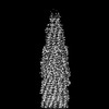
| Title | bottom segment of the bacteriophage M13 mini variant | |||||||||
 Components Components |
| |||||||||
 Keywords Keywords | VIRAL PROTEIN / M13 | |||||||||
| Function / homology |  Function and homology information Function and homology informationviral extrusion / virion attachment to host cell pilus / adhesion receptor-mediated virion attachment to host cell / helical viral capsid / host cell membrane / virion component / viral capsid / entry receptor-mediated virion attachment to host cell / symbiont entry into host cell / host cell plasma membrane / membrane Similarity search - Function | |||||||||
| Biological species |  Enterobacteria phage M13 (virus) Enterobacteria phage M13 (virus) | |||||||||
| Method | ELECTRON MICROSCOPY / single particle reconstruction / cryo EM / Resolution: 3.3 Å | |||||||||
 Authors Authors | Xiang, Y. / Jia, Q. | |||||||||
| Funding support |  China, 2items China, 2items
| |||||||||
 Citation Citation | Journal: To Be Published Title: Cryo-EM structure of a bacteriophage M13 mini variant Authors: Xiang, Y. / Jia, Q. | |||||||||
| History |
|
- Structure visualization
| Structure viewer | Molecule: MolmilJmol/JSmol |
|---|
- Downloads & links
-Download
| PDBx/mmCIF format | 8jwx.cif.gz | 338 KB | Display | PDBx/mmCIF format |
|---|---|---|---|---|
| PDB format | pdb8jwx.ent.gz | 272.6 KB | Display | PDB format |
| PDBx/mmJSON format | 8jwx.json.gz | Tree view | PDBx/mmJSON format | |
| Others |  Other downloads Other downloads |
-Validation report
| Arichive directory | https://data.pdbj.org/pub/pdb/validation_reports/jw/8jwxftp://data.pdbj.org/pub/pdb/validation_reports/jw/8jwx | HTTPS FTP |
|---|
-Related structure data
| Related structure data | 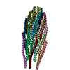 8jwwC M: map data used to model this data C: citing same article ( |
|---|---|
| Similar structure data |
-Links
 PDBj
PDBj- Assembly
| Deposited unit | 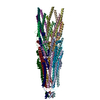
|
|---|---|
| 1 |
|
-Components
| #1: Protein | Mass: 42573.766 Da / Num. of mol.: 5 Source method: isolated from a genetically manipulated source Source: (gene. exp.) Enterobacteria phage M13 (virus) / Gene: III / Production host:  #2: Protein/peptide | Mass: 5243.014 Da / Num. of mol.: 15 Source method: isolated from a genetically manipulated source Source: (gene. exp.) Enterobacteria phage M13 (virus) / Gene: VIII / Production host: #3: Protein | Mass: 12357.984 Da / Num. of mol.: 5 Source method: isolated from a genetically manipulated source Source: (gene. exp.) Enterobacteria phage M13 (virus) / Gene: VI / Production host: |
|---|
-Experimental details
-Experiment
| Experiment | Method: ELECTRON MICROSCOPY |
|---|---|
| EM experiment | Aggregation state: PARTICLE / 3D reconstruction method: single particle reconstruction |
- Sample preparation
| Component | Name: bottom segment of the bacteriophage M13 mini variant / Type: COMPLEX / Entity ID: all / Source: NATURAL |
|---|---|
| Source (natural) | Organism: Enterobacteria phage M13 (virus) |
| Buffer solution | pH: 8 |
| Specimen | Embedding applied: NO / Shadowing applied: NO / Staining applied: NO / Vitrification applied: YES |
| Vitrification | Cryogen name: ETHANE |
- Electron microscopy imaging
| Experimental equipment |  Model: Titan Krios / Image courtesy: FEI Company |
|---|---|
| Microscopy | Model: FEI TITAN KRIOS |
| Electron gun | Electron source:  FIELD EMISSION GUN / Accelerating voltage: 300 kV / Illumination mode: FLOOD BEAM FIELD EMISSION GUN / Accelerating voltage: 300 kV / Illumination mode: FLOOD BEAM |
| Electron lens | Mode: BRIGHT FIELD / Nominal defocus max: 1700 nm / Nominal defocus min: 1200 nm |
| Image recording | Electron dose: 40 e/Å2 / Film or detector model: GATAN K3 (6k x 4k) |
- Processing
| CTF correction | Type: NONE |
|---|---|
| 3D reconstruction | Resolution: 3.3 Å / Resolution method: FSC 0.143 CUT-OFF / Num. of particles: 45177 / Symmetry type: POINT |
