 Movie
Movie Controller


+ Open data

- Basic information
| Entry | Database: PDB / ID: 8jv2 | ||||||
|---|---|---|---|---|---|---|---|
| Title | Structure of the SAR11 PotD in complex with proline | ||||||
 Components Components | Spermidine/putrescine-binding periplasmic protein | ||||||
 Keywords Keywords | TRANSPORT PROTEIN / PotD / substrate binding protein / solute-binding protein / periplasmic binding protein / ABC transporter system receptor / osmolyte / compatible solute / betaine / DMSP / GABA / amino acid / SAR11 / Candidatus Pelagibacter sp. HTCC7211 | ||||||
| Function / homology | Bacterial extracellular solute-binding protein / Bacterial extracellular solute-binding protein / periplasmic space / PROLINE / Spermidine/putrescine-binding periplasmic protein Function and homology information Function and homology information | ||||||
| Biological species | Pelagibacter sp. | ||||||
| Method |  X-RAY DIFFRACTION / SYNCHROTRON / MOLECULAR REPLACEMENT / Resolution: 1.3 Å X-RAY DIFFRACTION / SYNCHROTRON / MOLECULAR REPLACEMENT / Resolution: 1.3 Å | ||||||
 Authors Authors | Ma, Q. / Liu, C. | ||||||
| Funding support |  China, 1items China, 1items
| ||||||
 Citation Citation | Journal: To Be Published Title: Structure of the SAR11 PotD in complex with proline Authors: Ma, Q. / Liu, C. | ||||||
| History |
|
- Structure visualization
| Structure viewer | Molecule: MolmilJmol/JSmol |
|---|
- Downloads & links
-Download
| PDBx/mmCIF format | 8jv2.cif.gz | 173.9 KB | Display | PDBx/mmCIF format |
|---|---|---|---|---|
| PDB format | pdb8jv2.ent.gz | 134.9 KB | Display | PDB format |
| PDBx/mmJSON format | 8jv2.json.gz | Tree view | PDBx/mmJSON format | |
| Others |  Other downloads Other downloads |
-Validation report
| Summary document | 8jv2_validation.pdf.gz | 750.3 KB | Display | wwPDB validaton report |
|---|---|---|---|---|
| Full document | 8jv2_full_validation.pdf.gz | 750.5 KB | Display | |
| Data in XML | 8jv2_validation.xml.gz | 18.6 KB | Display | |
| Data in CIF | 8jv2_validation.cif.gz | 29.4 KB | Display | |
| Arichive directory | https://data.pdbj.org/pub/pdb/validation_reports/jv/8jv2ftp://data.pdbj.org/pub/pdb/validation_reports/jv/8jv2 | HTTPS FTP |
-Related structure data
| Similar structure data |
|---|
-Links
 PDBj
PDBj


- Assembly
| Deposited unit | 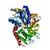
| ||||||||
|---|---|---|---|---|---|---|---|---|---|
| 1 |
| ||||||||
| Unit cell |
|
-Components
-Protein , 1 types, 1 molecules A
| #1: Protein | Mass: 39442.609 Da / Num. of mol.: 1 Source method: isolated from a genetically manipulated source Source: (gene. exp.)  Pelagibacter sp. (strain HTCC7211) (bacteria) Pelagibacter sp. (strain HTCC7211) (bacteria)Gene: potD, PB7211_697 / Production host: |
|---|
-Non-polymers , 5 types, 440 molecules 


| #2: Chemical | ChemComp-PRO /  Type: L-peptide linking / Mass: 115.130 Da / Num. of mol.: 1 / Source method: obtained synthetically / Formula: C5H9NO2 / Feature type: SUBJECT OF INVESTIGATION Type: L-peptide linking / Mass: 115.130 Da / Num. of mol.: 1 / Source method: obtained synthetically / Formula: C5H9NO2 / Feature type: SUBJECT OF INVESTIGATION | ||||||
|---|---|---|---|---|---|---|---|
| #3: Chemical |  Mass: 195.237 Da / Num. of mol.: 2 / Source method: obtained synthetically / Formula: C6H13NO4S / Comment: pH buffer*YM Mass: 195.237 Da / Num. of mol.: 2 / Source method: obtained synthetically / Formula: C6H13NO4S / Comment: pH buffer*YM#4: Chemical | ChemComp-PG4 / |  Mass: 194.226 Da / Num. of mol.: 1 / Source method: obtained synthetically / Formula: C8H18O5 / Comment: precipitant*YM Mass: 194.226 Da / Num. of mol.: 1 / Source method: obtained synthetically / Formula: C8H18O5 / Comment: precipitant*YM#5: Chemical |  Mass: 92.094 Da / Num. of mol.: 2 / Source method: isolated from a natural source / Formula: C3H8O3 Mass: 92.094 Da / Num. of mol.: 2 / Source method: isolated from a natural source / Formula: C3H8O3#6: Water | ChemComp-HOH / | Mass: 18.015 Da / Num. of mol.: 434 / Source method: isolated from a natural source / Formula: H2O |
-Details
| Has ligand of interest | Y |
|---|---|
| Has protein modification | Y |
-Experimental details
-Experiment
| Experiment | Method: X-RAY DIFFRACTION / Number of used crystals: 1 |
|---|
- Sample preparation
| Crystal | Density Matthews: 2.75 Å3/Da / Density % sol: 55.32 % |
|---|---|
| Crystal grow | Temperature: 293 K / Method: vapor diffusion, sitting drop Details: The protein in complex with L-proline was crystallized in drops containing 1 ul protein solution (60 mg/ml protein in buffer containing 10 mM HEPES, 150 mM NaCl, 50 mM L-proline, pH 7.5) and ...Details: The protein in complex with L-proline was crystallized in drops containing 1 ul protein solution (60 mg/ml protein in buffer containing 10 mM HEPES, 150 mM NaCl, 50 mM L-proline, pH 7.5) and 1 ul reservoir solution (100 mM MES pH 6.5, 20% w/v mPEG 550). |
-Data collection
| Diffraction | Mean temperature: 100 K / Serial crystal experiment: N |
|---|---|
| Diffraction source | Source: SYNCHROTRON / Site: SSRF / Beamline: BL19U1 / Wavelength: 0.97853 Å |
| Detector | Type: DECTRIS PILATUS3 6M / Detector: PIXEL / Date: Apr 6, 2023 |
| Radiation | Protocol: SINGLE WAVELENGTH / Monochromatic (M) / Laue (L): M / Scattering type: x-ray |
| Radiation wavelength | Wavelength: 0.97853 Å / Relative weight: 1 |
| Reflection | Resolution: 1.3→55.733 Å / Num. obs: 104876 / % possible obs: 97.7 % / Redundancy: 12.4 % / CC1/2: 1 / Rpim(I) all: 0.011 / Rrim(I) all: 0.041 / Rsym value: 0.039 / Net I/σ(I): 31.9 |
| Reflection shell | Resolution: 1.3→1.322 Å / Redundancy: 7.8 % / Mean I/σ(I) obs: 3.3 / Num. unique obs: 4306 / CC1/2: 0.874 / Rpim(I) all: 0.157 / Rrim(I) all: 0.457 / Rsym value: 0.426 / % possible all: 81 |
- Processing
| Software |
| |||||||||||||||||||||||||||||||||||||||||||||||||||||||||||||||||||||||||||||||||||||||||||||||||||||||||||||||||||||||||||||||||||||||||||||||||||||||||||||||||||||||||||||||||||||||||||||||||||||||||||||||||||||||||
|---|---|---|---|---|---|---|---|---|---|---|---|---|---|---|---|---|---|---|---|---|---|---|---|---|---|---|---|---|---|---|---|---|---|---|---|---|---|---|---|---|---|---|---|---|---|---|---|---|---|---|---|---|---|---|---|---|---|---|---|---|---|---|---|---|---|---|---|---|---|---|---|---|---|---|---|---|---|---|---|---|---|---|---|---|---|---|---|---|---|---|---|---|---|---|---|---|---|---|---|---|---|---|---|---|---|---|---|---|---|---|---|---|---|---|---|---|---|---|---|---|---|---|---|---|---|---|---|---|---|---|---|---|---|---|---|---|---|---|---|---|---|---|---|---|---|---|---|---|---|---|---|---|---|---|---|---|---|---|---|---|---|---|---|---|---|---|---|---|---|---|---|---|---|---|---|---|---|---|---|---|---|---|---|---|---|---|---|---|---|---|---|---|---|---|---|---|---|---|---|---|---|---|---|---|---|---|---|---|---|---|---|---|---|---|---|---|---|---|
| Refinement | Method to determine structure: MOLECULAR REPLACEMENT / Resolution: 1.3→26.235 Å / SU ML: 0.09 / Cross valid method: THROUGHOUT / σ(F): 1.36 / Phase error: 10.75 / Stereochemistry target values: ML
| |||||||||||||||||||||||||||||||||||||||||||||||||||||||||||||||||||||||||||||||||||||||||||||||||||||||||||||||||||||||||||||||||||||||||||||||||||||||||||||||||||||||||||||||||||||||||||||||||||||||||||||||||||||||||
| Solvent computation | Shrinkage radii: 0.9 Å / VDW probe radii: 1.11 Å / Solvent model: FLAT BULK SOLVENT MODEL | |||||||||||||||||||||||||||||||||||||||||||||||||||||||||||||||||||||||||||||||||||||||||||||||||||||||||||||||||||||||||||||||||||||||||||||||||||||||||||||||||||||||||||||||||||||||||||||||||||||||||||||||||||||||||
| Refinement step | Cycle: LAST / Resolution: 1.3→26.235 Å
| |||||||||||||||||||||||||||||||||||||||||||||||||||||||||||||||||||||||||||||||||||||||||||||||||||||||||||||||||||||||||||||||||||||||||||||||||||||||||||||||||||||||||||||||||||||||||||||||||||||||||||||||||||||||||
| Refine LS restraints |
| |||||||||||||||||||||||||||||||||||||||||||||||||||||||||||||||||||||||||||||||||||||||||||||||||||||||||||||||||||||||||||||||||||||||||||||||||||||||||||||||||||||||||||||||||||||||||||||||||||||||||||||||||||||||||
| LS refinement shell |
|
