- PDB-8jv0: Crystal structure of the SLA-2*1001 allele and ASFV antigenic pep... -
+
Open data
ID or keywords:
Loading...
-
Basic information
Entry
Database: PDB / ID: 8jv0
Title
Crystal structure of the SLA-2*1001 allele and ASFV antigenic peptide at 2.2A resolution
Components
Beta-2-microglobulin
MHC class I antigen
TYR-MET-ASN-CYS-SER-LEU-PRO-THR-TYR
Keywords
IMMUNE SYSTEM / SLA / MHC I / IMMUNOLOGY ANTIGEN
Function / homology
Function and homology information
ER-Phagosome pathway / Endosomal/Vacuolar pathway / DAP12 interactions / DAP12 signaling / Antigen Presentation: Folding, assembly and peptide loading of class I MHC / Immunoregulatory interactions between a Lymphoid and a non-Lymphoid cell / Neutrophil degranulation / antigen processing and presentation of peptide antigen via MHC class I / peptide antigen assembly with MHC class II protein complex / lumenal side of endoplasmic reticulum membrane ...ER-Phagosome pathway / Endosomal/Vacuolar pathway / DAP12 interactions / DAP12 signaling / Antigen Presentation: Folding, assembly and peptide loading of class I MHC / Immunoregulatory interactions between a Lymphoid and a non-Lymphoid cell / Neutrophil degranulation / antigen processing and presentation of peptide antigen via MHC class I / peptide antigen assembly with MHC class II protein complex / lumenal side of endoplasmic reticulum membrane / MHC class II protein complex / antigen processing and presentation of exogenous peptide antigen via MHC class II / positive regulation of immune response / MHC class I protein complex / peptide antigen binding / positive regulation of T cell activation / phagocytic vesicle membrane / MHC class II protein complex binding / late endosome membrane / immune response / lysosomal membrane / extracellular region Similarity search - Function
MHC class I, alpha chain, C-terminal / MHC_I C-terminus / MHC class I alpha chain, alpha1 alpha2 domains / Class I Histocompatibility antigen, domains alpha 1 and 2 / Beta-2-Microglobulin / : / MHC class I-like antigen recognition-like / MHC class I-like antigen recognition-like superfamily / MHC classes I/II-like antigen recognition protein / : ...MHC class I, alpha chain, C-terminal / MHC_I C-terminus / MHC class I alpha chain, alpha1 alpha2 domains / Class I Histocompatibility antigen, domains alpha 1 and 2 / Beta-2-Microglobulin / : / MHC class I-like antigen recognition-like / MHC class I-like antigen recognition-like superfamily / MHC classes I/II-like antigen recognition protein / : / Immunoglobulin/major histocompatibility complex, conserved site / Immunoglobulins and major histocompatibility complex proteins signature. / Immunoglobulin C-Type / Immunoglobulin C1-set / Immunoglobulin C1-set domain / Ig-like domain profile. / Immunoglobulin-like domain / Immunoglobulin-like domain superfamily / Immunoglobulin-like fold Similarity search - Domain/homology
A: MHC class I antigen B: Beta-2-microglobulin C: TYR-MET-ASN-CYS-SER-LEU-PRO-THR-TYR D: MHC class I antigen E: Beta-2-microglobulin F: TYR-MET-ASN-CYS-SER-LEU-PRO-THR-TYR
Mass: 18.015 Da / Num. of mol.: 109 / Source method: isolated from a natural source / Formula: H2O
Has protein modification
Y
-
Experimental details
-
Experiment
Experiment
Method: X-RAY DIFFRACTION / Number of used crystals: 1
-
Sample preparation
Crystal
Density Matthews: 2.76 Å3/Da / Density % sol: 55.49 %
Crystal grow
Temperature: 291 K / Method: vapor diffusion, sitting drop Details: 0.2 M Lithium sulfate monohydrate,0.1 M TRIS hydrochloride pH 8.5,30% w/v Polyethylene glycol 4,000
-
Data collection
Diffraction
Mean temperature: 100 K / Serial crystal experiment: N
 Movie
Movie Controller

 Yorodumi
 Open data
 Basic information
 Components
 Keywords
 Function and homology information
 Asfivirus
 X-RAY DIFFRACTION /
  Authors
 China, 1items
  Citation
 Structure visualization
 Downloads & links
 Other downloads
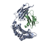
 PDBj
PDBj

 Assembly


 Mass: 18.015 Da / Num. of mol.: 109 / Source method: isolated from a natural source / Formula: H2O
 Sample preparation
 Processing