[English] 日本語
Yorodumi
- PDB-8jue: Crystal structure of glutaminase C in complex with compound 11 -

+
Open data


ID or keywords:

Loading...

-
Basic information

Entry
Database: PDB / ID: 8jue
TitleCrystal structure of glutaminase C in complex with compound 11
ComponentsGlutaminase kidney isoform, mitochondrial
KeywordsHYDROLASE/HYDROLASE INHIBITOR / Inhibitor / Complex / ANTITUMOR PROTEIN / HYDROLASE-HYDROLASE INHIBITOR complex
Function / homology
Function and homology information


glutamine catabolic process / regulation of respiratory gaseous exchange by nervous system process / glutamate biosynthetic process / Glutamate and glutamine metabolism / intracellular glutamate homeostasis / Glutamate Neurotransmitter Release Cycle / glutaminase / glutaminase activity / suckling behavior / TP53 Regulates Metabolic Genes ...glutamine catabolic process / regulation of respiratory gaseous exchange by nervous system process / glutamate biosynthetic process / Glutamate and glutamine metabolism / intracellular glutamate homeostasis / Glutamate Neurotransmitter Release Cycle / glutaminase / glutaminase activity / suckling behavior / TP53 Regulates Metabolic Genes / chemical synaptic transmission / protein homotetramerization / mitochondrial matrix / synapse / mitochondrion / cytosol
Similarity search - Function
Glutaminase, EF-hand domain / EF-hand domain / Glutaminase / Glutaminase / Ankyrin repeats (3 copies) / Ankyrin repeat profile. / Ankyrin repeat region circular profile. / ankyrin repeats / Ankyrin repeat / Ankyrin repeat-containing domain superfamily / Beta-lactamase/transpeptidase-like
Similarity search - Domain/homology
Chem-V59 / Glutaminase kidney isoform, mitochondrial
Similarity search - Component
Biological speciesHomo sapiens (human)
MethodX-RAY DIFFRACTION / SYNCHROTRON / MAD / Resolution: 2.39 Å
AuthorsWang, X. / Hanyu, S. / Tingting, D.
Funding support United Kingdom, 1items
OrganizationGrant numberCountry
CAMS Innovation Fund for Medical Sciences (CIFMS)CIFMS, No. 2021-I2M-1-028 United Kingdom
CitationJournal: Acs Med.Chem.Lett. / Year: 2023
Title: Targeting the Subpocket Enables the Discovery of Thiadiazole-Pyridazine Derivatives as Glutaminase C Inhibitors.
Authors: Sun, H. / Du, T. / Yang, M. / Liu, X. / Xue, X. / Chen, K. / Lang, X. / Chen, X. / Wang, B. / Wang, X.
History
DepositionJun 26, 2023Deposition site: PDBJ / Processing site: PDBJ
Revision 1.0Oct 11, 2023Provider: repository / Type: Initial release
Revision 1.1Nov 1, 2023Group: Database references / Category: citation / citation_author
Item: _citation.journal_volume / _citation.page_first ..._citation.journal_volume / _citation.page_first / _citation.page_last / _citation.pdbx_database_id_PubMed / _citation.title / _citation_author.identifier_ORCID

-
Structure visualization

Structure viewerMolecule:
MolmilJmol/JSmol

Downloads & links

-
Assembly

Deposited unit
A: Glutaminase kidney isoform, mitochondrial
B: Glutaminase kidney isoform, mitochondrial
C: Glutaminase kidney isoform, mitochondrial
D: Glutaminase kidney isoform, mitochondrial
hetero molecules


Theoretical massNumber of molelcules
Total (without water)236,0716
Polymers235,1234
Non-polymers9472
Water6,485360
1


  • Idetical with deposited unit
  • defined by author&software
  • Evidence: native gel electrophoresis
TypeNameSymmetry operationNumber
identity operation1_555x,y,z1
Buried area11430 Å2
ΔGint-47 kcal/mol
Surface area58780 Å2
MethodPISA
Unit cell
Length a, b, c (Å)49.649, 139.510, 178.094
Angle α, β, γ (deg.)90.00, 93.49, 90.00
Int Tables number4
Space group name H-MP1211

-
Components

#1: Protein
Glutaminase kidney isoform, mitochondrial / GLS / K-glutaminase / L-glutamine amidohydrolase


Mass: 58780.859 Da / Num. of mol.: 4
Source method: isolated from a genetically manipulated source
Source: (gene. exp.) Homo sapiens (human) / Gene: GLS, GLS1, KIAA0838 / Production host: Escherichia coli BL21(DE3) (bacteria) / References: UniProt: O94925, glutaminase
#2: Chemical ChemComp-V59 / 2-(3-phenoxyphenyl)-N-[5-[[(3R)-1-pyridazin-3-ylpyrrolidin-3-yl]amino]-1,3,4-thiadiazol-2-yl]ethanamide


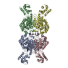
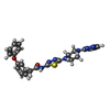
Mass: 473.550 Da / Num. of mol.: 2 / Source method: obtained synthetically / Formula: C24H23N7O2S
#3: Water ChemComp-HOH / water


Mass: 18.015 Da / Num. of mol.: 360 / Source method: isolated from a natural source / Formula: H2O
Has ligand of interestY

-
Experimental details

-
Experiment

ExperimentMethod: X-RAY DIFFRACTION / Number of used crystals: 1

-
Sample preparation

CrystalDensity Matthews: 2.47 Å3/Da / Density % sol: 50.19 %
Crystal growTemperature: 293 K / Method: vapor diffusion, hanging drop
Details: 18% Polyethylene glycol 3350, 0.2 M magnesium chloride, 0.08 M 2-[4-(2-Hydroxyethyl)-1-piperazinyl]ethanesulfonicacid pH 7.5

-
Data collection

DiffractionMean temperature: 100 K / Serial crystal experiment: N
Diffraction sourceSource: SYNCHROTRON / Site: SSRF / Beamline: BL02U1 / Wavelength: 0.979 Å
DetectorType: DECTRIS EIGER2 S 9M / Detector: PIXEL / Date: Jun 10, 2023
RadiationProtocol: SINGLE WAVELENGTH / Monochromatic (M) / Laue (L): M / Scattering type: x-ray
Radiation wavelengthWavelength: 0.979 Å / Relative weight: 1
ReflectionResolution: 2.39→109.99 Å / Num. obs: 89723 / % possible obs: 94.39 % / Redundancy: 5.7 % / CC1/2: 0.998 / Rmerge(I) obs: 0.05677 / Rpim(I) all: 0.02589 / Net I/σ(I): 22.85
Reflection shellResolution: 2.39→2.475 Å / Rmerge(I) obs: 0.2157 / Mean I/σ(I) obs: 8.65 / Num. unique obs: 9510 / CC1/2: 0.979 / Rpim(I) all: 0.09593 / Rrim(I) all: 0.2365

-
Processing

Software
NameVersionClassification
REFMAC5.8.0352refinement
XDSdata reduction
XDSdata scaling
PHENIXphasing
RefinementMethod to determine structure: MAD / Resolution: 2.39→109.99 Å / Cor.coef. Fo:Fc: 0.952 / Cor.coef. Fo:Fc free: 0.919 / SU B: 7.699 / SU ML: 0.174 / Cross valid method: THROUGHOUT / ESU R: 0.294 / ESU R Free: 0.238 / Stereochemistry target values: MAXIMUM LIKELIHOOD / Details: HYDROGENS HAVE BEEN ADDED IN THE RIDING POSITIONS
RfactorNum. reflection% reflectionSelection details
Rfree0.24408 4490 5.1 %RANDOM
Rwork0.18875 ---
obs0.1915 84303 92.94 %-
Solvent computationIon probe radii: 0.8 Å / Shrinkage radii: 0.8 Å / VDW probe radii: 1.2 Å / Solvent model: MASK
Displacement parametersBiso mean: 45.974 Å2
Baniso -1Baniso -2Baniso -3
1-3.31 Å2-0 Å2-0.73 Å2
2---2.18 Å20 Å2
3----1.03 Å2
Refinement stepCycle: 1 / Resolution: 2.39→109.99 Å
ProteinNucleic acidLigandSolventTotal
Num. atoms12436 0 68 360 12864
Refine LS restraints
Refine-IDTypeDev idealDev ideal targetNumber
X-RAY DIFFRACTIONr_bond_refined_d0.0070.01212784
X-RAY DIFFRACTIONr_bond_other_d0.0010.01611751
X-RAY DIFFRACTIONr_angle_refined_deg1.4821.64717241
X-RAY DIFFRACTIONr_angle_other_deg0.4871.56727421
X-RAY DIFFRACTIONr_dihedral_angle_1_deg7.15751577
X-RAY DIFFRACTIONr_dihedral_angle_2_deg8.151560
X-RAY DIFFRACTIONr_dihedral_angle_3_deg17.023102230
X-RAY DIFFRACTIONr_dihedral_angle_4_deg
X-RAY DIFFRACTIONr_chiral_restr0.0680.21892
X-RAY DIFFRACTIONr_gen_planes_refined0.0060.0214369
X-RAY DIFFRACTIONr_gen_planes_other0.0010.022571
X-RAY DIFFRACTIONr_nbd_refined
X-RAY DIFFRACTIONr_nbd_other
X-RAY DIFFRACTIONr_nbtor_refined
X-RAY DIFFRACTIONr_nbtor_other
X-RAY DIFFRACTIONr_xyhbond_nbd_refined
X-RAY DIFFRACTIONr_xyhbond_nbd_other
X-RAY DIFFRACTIONr_metal_ion_refined
X-RAY DIFFRACTIONr_metal_ion_other
X-RAY DIFFRACTIONr_symmetry_vdw_refined
X-RAY DIFFRACTIONr_symmetry_vdw_other
X-RAY DIFFRACTIONr_symmetry_hbond_refined
X-RAY DIFFRACTIONr_symmetry_hbond_other
X-RAY DIFFRACTIONr_symmetry_metal_ion_refined
X-RAY DIFFRACTIONr_symmetry_metal_ion_other
X-RAY DIFFRACTIONr_mcbond_it3.5434.4986344
X-RAY DIFFRACTIONr_mcbond_other3.5414.4986344
X-RAY DIFFRACTIONr_mcangle_it5.3636.7267909
X-RAY DIFFRACTIONr_mcangle_other5.3636.7277910
X-RAY DIFFRACTIONr_scbond_it4.3055.0836440
X-RAY DIFFRACTIONr_scbond_other4.3055.0846441
X-RAY DIFFRACTIONr_scangle_it
X-RAY DIFFRACTIONr_scangle_other6.5627.4099333
X-RAY DIFFRACTIONr_long_range_B_refined8.8853.7614095
X-RAY DIFFRACTIONr_long_range_B_other8.88353.74614046
X-RAY DIFFRACTIONr_rigid_bond_restr
X-RAY DIFFRACTIONr_sphericity_free
X-RAY DIFFRACTIONr_sphericity_bonded
LS refinement shellResolution: 2.391→2.453 Å / Total num. of bins used: 20
RfactorNum. reflection% reflection
Rfree0.315 364 -
Rwork0.268 6667 -
obs--99.84 %

+
About Yorodumi

-
News

-
Feb 9, 2022. New format data for meta-information of EMDB entries

New format data for meta-information of EMDB entries

  • Version 3 of the EMDB header file is now the official format.
  • The previous official version 1.9 will be removed from the archive.

Related info.:EMDB header

External links:wwPDB to switch to version 3 of the EMDB data model

-
Aug 12, 2020. Covid-19 info

Covid-19 info

URL: https://pdbjlvh1.pdbj.org/emnavi/covid19.php

New page: Covid-19 featured information page in EM Navigator.

Related info.:Covid-19 info / Mar 5, 2020. Novel coronavirus structure data

+
Mar 5, 2020. Novel coronavirus structure data

Novel coronavirus structure data

Related info.:Yorodumi Speices / Aug 12, 2020. Covid-19 info

External links:COVID-19 featured content - PDBj / Molecule of the Month (242):Coronavirus Proteases

+
Jan 31, 2019. EMDB accession codes are about to change! (news from PDBe EMDB page)

EMDB accession codes are about to change! (news from PDBe EMDB page)

  • The allocation of 4 digits for EMDB accession codes will soon come to an end. Whilst these codes will remain in use, new EMDB accession codes will include an additional digit and will expand incrementally as the available range of codes is exhausted. The current 4-digit format prefixed with “EMD-” (i.e. EMD-XXXX) will advance to a 5-digit format (i.e. EMD-XXXXX), and so on. It is currently estimated that the 4-digit codes will be depleted around Spring 2019, at which point the 5-digit format will come into force.
  • The EM Navigator/Yorodumi systems omit the EMD- prefix.

Related info.:Q: What is EMD? / ID/Accession-code notation in Yorodumi/EM Navigator

External links:EMDB Accession Codes are Changing Soon! / Contact to PDBj

+
Jul 12, 2017. Major update of PDB

Major update of PDB

  • wwPDB released updated PDB data conforming to the new PDBx/mmCIF dictionary.
  • This is a major update changing the version number from 4 to 5, and with Remediation, in which all the entries are updated.
  • In this update, many items about electron microscopy experimental information are reorganized (e.g. em_software).
  • Now, EM Navigator and Yorodumi are based on the updated data.

External links:wwPDB Remediation / Enriched Model Files Conforming to OneDep Data Standards Now Available in the PDB FTP Archive

-
Yorodumi

Thousand views of thousand structures

  • Yorodumi is a browser for structure data from EMDB, PDB, SASBDB, etc.
  • This page is also the successor to EM Navigator detail page, and also detail information page/front-end page for Omokage search.
  • The word "yorodu" (or yorozu) is an old Japanese word meaning "ten thousand". "mi" (miru) is to see.

Related info.:EMDB / PDB / SASBDB / Comparison of 3 databanks / Yorodumi Search / Aug 31, 2016. New EM Navigator & Yorodumi / Yorodumi Papers / Jmol/JSmol / Function and homology information / Changes in new EM Navigator and Yorodumi

Read more