 Movie
Movie Controller


+ Open data

- Basic information
| Entry | Database: PDB / ID: 8ju3 | ||||||
|---|---|---|---|---|---|---|---|
| Title | Mu phage tail fiber | ||||||
 Components Components | Tail fiber protein S,Tail fiber protein S' | ||||||
 Keywords Keywords | VIRAL PROTEIN / bacteriophage | ||||||
| Function / homology |  Function and homology information Function and homology informationviral tropism switching / virus tail, fiber / adhesion receptor-mediated virion attachment to host cell / host cell cytoplasm / symbiont entry into host cell Similarity search - Function | ||||||
| Biological species |  Escherichia phage Mu (virus) Escherichia phage Mu (virus) | ||||||
| Method |  X-RAY DIFFRACTION / SYNCHROTRON / MOLECULAR REPLACEMENT / Resolution: 2 Å X-RAY DIFFRACTION / SYNCHROTRON / MOLECULAR REPLACEMENT / Resolution: 2 Å | ||||||
 Authors Authors | Yamashita, E. / Takeda, S. | ||||||
| Funding support | 1items
| ||||||
 Citation Citation | Journal: Virology / Year: 2024 Title: Determination of the three-dimensional structure of bacteriophage Mu(-) tail fiber and its characterization. Authors: Mori, Y. / Yamashita, E. / Nakagawa, A. / Matsuzawa, T. / Inagaki, M. / Aiba, Y. / Tanaka, S. / Hatori, S. / Ayami, M. / Takeda, S. | ||||||
| History |
|
- Structure visualization
| Structure viewer | Molecule: MolmilJmol/JSmol |
|---|
- Downloads & links
-Download
| PDBx/mmCIF format | 8ju3.cif.gz | 165.3 KB | Display | PDBx/mmCIF format |
|---|---|---|---|---|
| PDB format | pdb8ju3.ent.gz | 106.5 KB | Display | PDB format |
| PDBx/mmJSON format | 8ju3.json.gz | Tree view | PDBx/mmJSON format | |
| Others |  Other downloads Other downloads |
-Validation report
| Summary document | 8ju3_validation.pdf.gz | 435 KB | Display | wwPDB validaton report |
|---|---|---|---|---|
| Full document | 8ju3_full_validation.pdf.gz | 443.8 KB | Display | |
| Data in XML | 8ju3_validation.xml.gz | 15.2 KB | Display | |
| Data in CIF | 8ju3_validation.cif.gz | 22.3 KB | Display | |
| Arichive directory | https://data.pdbj.org/pub/pdb/validation_reports/ju/8ju3ftp://data.pdbj.org/pub/pdb/validation_reports/ju/8ju3 | HTTPS FTP |
-Related structure data
| Similar structure data |
|---|
-Links
 PDBj
PDBj- Assembly
| Deposited unit | 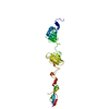
| ||||||||||||
|---|---|---|---|---|---|---|---|---|---|---|---|---|---|
| 1 | 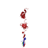
| ||||||||||||
| Unit cell |
| ||||||||||||
| Components on special symmetry positions |
|
-Components
| #1: Protein | Mass: 52697.312 Da / Num. of mol.: 1 Source method: isolated from a genetically manipulated source Source: (gene. exp.) Escherichia phage Mu (virus) / Gene: S, Mup49, S', Mup52Production host:  Variant (production host): pLysS / References: UniProt: Q9T1V0, UniProt: P0DJY6 |
|---|---|
| #2: Water | ChemComp-HOH /  Mass: 18.015 Da / Num. of mol.: 232 / Source method: isolated from a natural source / Formula: H2O Mass: 18.015 Da / Num. of mol.: 232 / Source method: isolated from a natural source / Formula: H2O |
-Experimental details
-Experiment
| Experiment | Method: X-RAY DIFFRACTION / Number of used crystals: 1 |
|---|
- Sample preparation
| Crystal | Density Matthews: 5.16 Å3/Da / Density % sol: 76.17 % |
|---|---|
| Crystal grow | Temperature: 293 K / Method: vapor diffusion, sitting drop / pH: 7.5 / Details: 0.1M HEPES (pH 7.5), 4.3M NaCl |
-Data collection
| Diffraction | Mean temperature: 100 K / Serial crystal experiment: N |
|---|---|
| Diffraction source | Source: SYNCHROTRON / Site: SPring-8  / Beamline: BL44XU / Wavelength: 0.9 Å / Beamline: BL44XU / Wavelength: 0.9 Å |
| Detector | Type: DECTRIS EIGER X 16M / Detector: PIXEL / Date: Oct 27, 2021 |
| Radiation | Protocol: SINGLE WAVELENGTH / Monochromatic (M) / Laue (L): M / Scattering type: x-ray |
| Radiation wavelength | Wavelength: 0.9 Å / Relative weight: 1 |
| Reflection | Resolution: 2→49.88 Å / Num. obs: 76418 / % possible obs: 99.75 % / Redundancy: 9.39 % / Biso Wilson estimate: 36.52 Å2 / CC1/2: 0.996 / Net I/σ(I): 8.4 |
| Reflection shell | Resolution: 2→2.03 Å / Num. unique obs: 3548 / CC1/2: 0.611 |
- Processing
| Software |
| ||||||||||||||||||||||||||||||||||||||||||||||||||||||||||||||||||||||||||||||||||||||||||||||||||||||||||||||||||||||||||||||||||||||||||||||||||||||||||||||||||||||||||||||||||||||||||||||||||||
|---|---|---|---|---|---|---|---|---|---|---|---|---|---|---|---|---|---|---|---|---|---|---|---|---|---|---|---|---|---|---|---|---|---|---|---|---|---|---|---|---|---|---|---|---|---|---|---|---|---|---|---|---|---|---|---|---|---|---|---|---|---|---|---|---|---|---|---|---|---|---|---|---|---|---|---|---|---|---|---|---|---|---|---|---|---|---|---|---|---|---|---|---|---|---|---|---|---|---|---|---|---|---|---|---|---|---|---|---|---|---|---|---|---|---|---|---|---|---|---|---|---|---|---|---|---|---|---|---|---|---|---|---|---|---|---|---|---|---|---|---|---|---|---|---|---|---|---|---|---|---|---|---|---|---|---|---|---|---|---|---|---|---|---|---|---|---|---|---|---|---|---|---|---|---|---|---|---|---|---|---|---|---|---|---|---|---|---|---|---|---|---|---|---|---|---|---|---|
| Refinement | Method to determine structure: MOLECULAR REPLACEMENT / Resolution: 2→49.88 Å / SU ML: 0.1994 / Cross valid method: FREE R-VALUE / σ(F): 1.34 / Phase error: 18.5094 Stereochemistry target values: GeoStd + Monomer Library + CDL v1.2
| ||||||||||||||||||||||||||||||||||||||||||||||||||||||||||||||||||||||||||||||||||||||||||||||||||||||||||||||||||||||||||||||||||||||||||||||||||||||||||||||||||||||||||||||||||||||||||||||||||||
| Solvent computation | Shrinkage radii: 0.9 Å / VDW probe radii: 1.11 Å / Solvent model: FLAT BULK SOLVENT MODEL | ||||||||||||||||||||||||||||||||||||||||||||||||||||||||||||||||||||||||||||||||||||||||||||||||||||||||||||||||||||||||||||||||||||||||||||||||||||||||||||||||||||||||||||||||||||||||||||||||||||
| Displacement parameters | Biso mean: 65.37 Å2 | ||||||||||||||||||||||||||||||||||||||||||||||||||||||||||||||||||||||||||||||||||||||||||||||||||||||||||||||||||||||||||||||||||||||||||||||||||||||||||||||||||||||||||||||||||||||||||||||||||||
| Refinement step | Cycle: LAST / Resolution: 2→49.88 Å
| ||||||||||||||||||||||||||||||||||||||||||||||||||||||||||||||||||||||||||||||||||||||||||||||||||||||||||||||||||||||||||||||||||||||||||||||||||||||||||||||||||||||||||||||||||||||||||||||||||||
| Refine LS restraints |
| ||||||||||||||||||||||||||||||||||||||||||||||||||||||||||||||||||||||||||||||||||||||||||||||||||||||||||||||||||||||||||||||||||||||||||||||||||||||||||||||||||||||||||||||||||||||||||||||||||||
| LS refinement shell |
| ||||||||||||||||||||||||||||||||||||||||||||||||||||||||||||||||||||||||||||||||||||||||||||||||||||||||||||||||||||||||||||||||||||||||||||||||||||||||||||||||||||||||||||||||||||||||||||||||||||
| Refinement TLS params. | Method: refined / Origin x: -30.6839202554 Å / Origin y: 10.5883412353 Å / Origin z: 33.6374153333 Å
| ||||||||||||||||||||||||||||||||||||||||||||||||||||||||||||||||||||||||||||||||||||||||||||||||||||||||||||||||||||||||||||||||||||||||||||||||||||||||||||||||||||||||||||||||||||||||||||||||||||
| Refinement TLS group | Selection details: all |
