[English] 日本語
Yorodumi
- PDB-8jtk: Structure of AYWB phytoplasma SAP05 recognizing AtRpn10 -

+
Open data


ID or keywords:

Loading...

-
Basic information

Entry
Database: PDB / ID: 8jtk
TitleStructure of AYWB phytoplasma SAP05 recognizing AtRpn10
Components
  • 26S proteasome non-ATPase regulatory subunit 4 homolog
  • Sequence-variable mosaic (SVM) signal sequence domain-containing protein
KeywordsPLANT PROTEIN / Complex / Proteasome / Hijack
Function / homology
Function and homology information


stamen formation / post-embryonic root development / response to cytokinin / root hair elongation / peptide receptor activity / regulation of seed germination / response to misfolded protein / leaf senescence / leaf development / pollen development ...stamen formation / post-embryonic root development / response to cytokinin / root hair elongation / peptide receptor activity / regulation of seed germination / response to misfolded protein / leaf senescence / leaf development / pollen development / response to auxin / response to sucrose / response to abscisic acid / proteasome regulatory particle, base subcomplex / proteasome assembly / polyubiquitin modification-dependent protein binding / response to salt stress / proteasome complex / protein catabolic process / response to heat / proteasome-mediated ubiquitin-dependent protein catabolic process / DNA damage response / nucleus / plasma membrane / cytosol
Similarity search - Function
Sequence-variable mosaic (SVM), signal sequence / SVM protein signal sequence / : / Ubiquitin interaction motif / Ubiquitin-interacting motif. / von Willebrand factor type A domain / Ubiquitin interacting motif / Ubiquitin-interacting motif (UIM) domain profile. / von Willebrand factor (vWF) type A domain / VWFA domain profile. ...Sequence-variable mosaic (SVM), signal sequence / SVM protein signal sequence / : / Ubiquitin interaction motif / Ubiquitin-interacting motif. / von Willebrand factor type A domain / Ubiquitin interacting motif / Ubiquitin-interacting motif (UIM) domain profile. / von Willebrand factor (vWF) type A domain / VWFA domain profile. / von Willebrand factor, type A / von Willebrand factor A-like domain superfamily
Similarity search - Domain/homology
26S proteasome non-ATPase regulatory subunit 4 homolog / Sequence-variable mosaic (SVM) signal sequence domain-containing protein
Similarity search - Component
Biological speciesArabidopsis thaliana (thale cress)
Aster yellows witches'-broom phytoplasma
MethodX-RAY DIFFRACTION / SYNCHROTRON / MOLECULAR REPLACEMENT / Resolution: 1.57 Å
AuthorsDu, Y.X. / Zhang, L.Y. / Zheng, Q.Y.
Funding support China, 1items
OrganizationGrant numberCountry
Other governmentYFC3401501 China
CitationJournal: To Be Published
Title: Structural basis for SAP05 driving ubiquitin-independent protein degradation
Authors: Zhang, L.Y. / Du, Y.X. / Zheng, Q.Y.
History
DepositionJun 22, 2023Deposition site: PDBJ / Processing site: PDBC
Revision 1.0Jul 19, 2023Provider: repository / Type: Initial release
Revision 1.1May 29, 2024Group: Data collection / Category: chem_comp_atom / chem_comp_bond

-
Structure visualization

Structure viewerMolecule:
MolmilJmol/JSmol

Downloads & links

-
Assembly

Deposited unit
B: 26S proteasome non-ATPase regulatory subunit 4 homolog
A: Sequence-variable mosaic (SVM) signal sequence domain-containing protein


Theoretical massNumber of molelcules
Total (without water)33,1912
Polymers33,1912
Non-polymers00
Water3,603200
1


  • Idetical with deposited unit
  • defined by author&software
  • Evidence: gel filtration
TypeNameSymmetry operationNumber
identity operation1_555x,y,z1
Buried area1610 Å2
ΔGint-8 kcal/mol
Surface area13980 Å2
MethodPISA
Unit cell
Length a, b, c (Å)49.240, 49.240, 215.154
Angle α, β, γ (deg.)90.000, 90.000, 120.000
Int Tables number154
Space group name H-MP3221
Space group name HallP322"
Symmetry operation#1: x,y,z
#2: -y,x-y,z+2/3
#3: -x+y,-x,z+1/3
#4: x-y,-y,-z+1/3
#5: -x,-x+y,-z+2/3
#6: y,x,-z
Components on special symmetry positions
IDModelComponents
11B-258-

HOH

-
Components

#1: Protein 26S proteasome non-ATPase regulatory subunit 4 homolog / 26S proteasome regulatory subunit RPN10 / AtRPN10 / 26S proteasome regulatory subunit S5A homolog / ...26S proteasome regulatory subunit RPN10 / AtRPN10 / 26S proteasome regulatory subunit S5A homolog / Multiubiquitin chain-binding protein 1 / AtMCB1


Mass: 20879.832 Da / Num. of mol.: 1
Source method: isolated from a genetically manipulated source
Source: (gene. exp.) Arabidopsis thaliana (thale cress) / Gene: RPN10, MBP1, MCB1, At4g38630, F20M13.190, T9A14.7 / Plasmid: pGEX-6p-1 / Production host: Escherichia coli (E. coli) / References: UniProt: P55034
#2: Protein Sequence-variable mosaic (SVM) signal sequence domain-containing protein


Mass: 12310.920 Da / Num. of mol.: 1
Source method: isolated from a genetically manipulated source
Source: (gene. exp.) Aster yellows witches'-broom phytoplasma (strain AYWB) (bacteria)
Gene: AYWB_032 / Plasmid: pET28a / Production host: Escherichia coli (E. coli) / References: UniProt: Q2NK94
#3: Water ChemComp-HOH / water


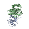
Mass: 18.015 Da / Num. of mol.: 200 / Source method: isolated from a natural source / Formula: H2O

-
Experimental details

-
Experiment

ExperimentMethod: X-RAY DIFFRACTION / Number of used crystals: 1

-
Sample preparation

CrystalDensity Matthews: 2.25 Å3/Da / Density % sol: 45.25 %
Crystal growTemperature: 291 K / Method: vapor diffusion, sitting drop
Details: 0.2 M Sodium malonate pH 5.0, 20% w/v Polyethylene glycol 3,350

-
Data collection

DiffractionMean temperature: 100 K / Serial crystal experiment: N
Diffraction sourceSource: SYNCHROTRON / Site: SSRF / Beamline: BL10U2 / Wavelength: 0.979183 Å
DetectorType: DECTRIS EIGER2 S 9M / Detector: PIXEL / Date: Dec 17, 2022
RadiationProtocol: SINGLE WAVELENGTH / Monochromatic (M) / Laue (L): M / Scattering type: x-ray
Radiation wavelengthWavelength: 0.979183 Å / Relative weight: 1
ReflectionResolution: 1.57→71.72 Å / Num. obs: 42242 / % possible obs: 95.9 % / Redundancy: 8.2 % / Biso Wilson estimate: 26.5 Å2 / CC1/2: 0.999 / Rmerge(I) obs: 0.056 / Rpim(I) all: 0.021 / Rrim(I) all: 0.06 / Net I/σ(I): 18
Reflection shellResolution: 1.57→1.65 Å / Rmerge(I) obs: 0.768 / Mean I/σ(I) obs: 2.6 / Num. unique obs: 6330 / CC1/2: 0.736 / Rpim(I) all: 0.314 / Rrim(I) all: 0.831 / % possible all: 100

-
Processing

Software
NameVersionClassification
PHENIX1.19.2_4158refinement
autoPROC1.0.5data processing
Aimless0.7.7data scaling
PHENIX1.19.2_4158phasing
RefinementMethod to determine structure: MOLECULAR REPLACEMENT / Resolution: 1.57→24.93 Å / SU ML: 0.1964 / Cross valid method: FREE R-VALUE / σ(F): 1.38 / Phase error: 22.9635
Stereochemistry target values: GeoStd + Monomer Library + CDL v1.2
RfactorNum. reflection% reflection
Rfree0.2154 2039 4.84 %
Rwork0.187 40103 -
obs0.1883 42142 95.86 %
Solvent computationShrinkage radii: 0.9 Å / VDW probe radii: 1.11 Å / Solvent model: FLAT BULK SOLVENT MODEL
Displacement parametersBiso mean: 38.6 Å2
Refinement stepCycle: LAST / Resolution: 1.57→24.93 Å
ProteinNucleic acidLigandSolventTotal
Num. atoms2322 0 0 200 2522
Refine LS restraints
Refine-IDTypeDev idealNumber
X-RAY DIFFRACTIONf_bond_d0.00762361
X-RAY DIFFRACTIONf_angle_d1.0333192
X-RAY DIFFRACTIONf_chiral_restr0.066361
X-RAY DIFFRACTIONf_plane_restr0.0086419
X-RAY DIFFRACTIONf_dihedral_angle_d15.8595887
LS refinement shell
Resolution (Å)Rfactor RfreeNum. reflection RfreeRfactor RworkNum. reflection RworkRefine-ID% reflection obs (%)
1.57-1.60.32121620.27722724X-RAY DIFFRACTION99.9
1.6-1.640.33141510.26792748X-RAY DIFFRACTION99.83
1.64-1.690.32031480.25662687X-RAY DIFFRACTION100
1.69-1.740.28961560.25422732X-RAY DIFFRACTION100
1.74-1.790.25591190.23742735X-RAY DIFFRACTION99.96
1.79-1.860.23561460.20952768X-RAY DIFFRACTION100
1.86-1.930.3215690.2151566X-RAY DIFFRACTION56.91
1.93-2.020.21691070.20852429X-RAY DIFFRACTION87.84
2.02-2.130.23241420.20522806X-RAY DIFFRACTION100
2.13-2.260.23861270.18782590X-RAY DIFFRACTION93.37
2.26-2.430.21721550.18582765X-RAY DIFFRACTION100
2.43-2.680.2281540.20122793X-RAY DIFFRACTION100
2.68-3.060.23041230.18952862X-RAY DIFFRACTION99.93
3.06-3.860.18961510.17492851X-RAY DIFFRACTION100
3.86-24.930.17711290.16113047X-RAY DIFFRACTION99.22

+
About Yorodumi

-
News

-
Feb 9, 2022. New format data for meta-information of EMDB entries

New format data for meta-information of EMDB entries

  • Version 3 of the EMDB header file is now the official format.
  • The previous official version 1.9 will be removed from the archive.

Related info.:EMDB header

External links:wwPDB to switch to version 3 of the EMDB data model

-
Aug 12, 2020. Covid-19 info

Covid-19 info

URL: https://pdbjlvh1.pdbj.org/emnavi/covid19.php

New page: Covid-19 featured information page in EM Navigator.

Related info.:Covid-19 info / Mar 5, 2020. Novel coronavirus structure data

+
Mar 5, 2020. Novel coronavirus structure data

Novel coronavirus structure data

Related info.:Yorodumi Speices / Aug 12, 2020. Covid-19 info

External links:COVID-19 featured content - PDBj / Molecule of the Month (242):Coronavirus Proteases

+
Jan 31, 2019. EMDB accession codes are about to change! (news from PDBe EMDB page)

EMDB accession codes are about to change! (news from PDBe EMDB page)

  • The allocation of 4 digits for EMDB accession codes will soon come to an end. Whilst these codes will remain in use, new EMDB accession codes will include an additional digit and will expand incrementally as the available range of codes is exhausted. The current 4-digit format prefixed with “EMD-” (i.e. EMD-XXXX) will advance to a 5-digit format (i.e. EMD-XXXXX), and so on. It is currently estimated that the 4-digit codes will be depleted around Spring 2019, at which point the 5-digit format will come into force.
  • The EM Navigator/Yorodumi systems omit the EMD- prefix.

Related info.:Q: What is EMD? / ID/Accession-code notation in Yorodumi/EM Navigator

External links:EMDB Accession Codes are Changing Soon! / Contact to PDBj

+
Jul 12, 2017. Major update of PDB

Major update of PDB

  • wwPDB released updated PDB data conforming to the new PDBx/mmCIF dictionary.
  • This is a major update changing the version number from 4 to 5, and with Remediation, in which all the entries are updated.
  • In this update, many items about electron microscopy experimental information are reorganized (e.g. em_software).
  • Now, EM Navigator and Yorodumi are based on the updated data.

External links:wwPDB Remediation / Enriched Model Files Conforming to OneDep Data Standards Now Available in the PDB FTP Archive

-
Yorodumi

Thousand views of thousand structures

  • Yorodumi is a browser for structure data from EMDB, PDB, SASBDB, etc.
  • This page is also the successor to EM Navigator detail page, and also detail information page/front-end page for Omokage search.
  • The word "yorodu" (or yorozu) is an old Japanese word meaning "ten thousand". "mi" (miru) is to see.

Related info.:EMDB / PDB / SASBDB / Comparison of 3 databanks / Yorodumi Search / Aug 31, 2016. New EM Navigator & Yorodumi / Yorodumi Papers / Jmol/JSmol / Function and homology information / Changes in new EM Navigator and Yorodumi

Read more