 ムービー
ムービー コントローラー


+ データを開く

- 基本情報
基本情報
| 登録情報 | データベース: PDB / ID: 8jqz | ||||||
|---|---|---|---|---|---|---|---|
| タイトル | Crystal Structure of GppNHp-bound mIRGB10 | ||||||
 要素 要素 | Immunity-related GTPase family member b10 | ||||||
 キーワード キーワード | IMMUNE SYSTEM / GTP hydrolase | ||||||
| 機能・相同性 |  機能・相同性情報 機能・相同性情報autophagosome assembly / cellular response to interferon-beta / innate immune response / GTPase activity / endoplasmic reticulum membrane / GTP binding 類似検索 - 分子機能 | ||||||
| 生物種 |  | ||||||
| 手法 |  X線回折 / シンクロトロン / 分子置換 / 解像度: 3.05 Å X線回折 / シンクロトロン / 分子置換 / 解像度: 3.05 Å | ||||||
 データ登録者 データ登録者 | Ha, H.J. / Park, H.H. | ||||||
| 資金援助 |  韓国, 1件 韓国, 1件
| ||||||
 引用 引用 | ジャーナル: Front Immunol / 年: 2023 タイトル: Structural basis of IRGB10 oligomerization by GTP hydrolysis. 著者: Ha, H.J. / Kim, J.H. / Lee, G.H. / Kim, S. / Park, H.H. | ||||||
| 履歴 |
|
- 構造の表示
構造の表示
| 構造ビューア | 分子: MolmilJmol/JSmol |
|---|
- ダウンロードとリンク
-ダウンロード
| PDBx/mmCIF形式 | 8jqz.cif.gz | 376.3 KB | 表示 | PDBx/mmCIF形式 |
|---|---|---|---|---|
| PDB形式 | pdb8jqz.ent.gz | 260.7 KB | 表示 | PDB形式 |
| PDBx/mmJSON形式 | 8jqz.json.gz | ツリー表示 | PDBx/mmJSON形式 | |
| その他 |  その他のダウンロード その他のダウンロード |
-検証レポート
| 文書・要旨 | 8jqz_validation.pdf.gz | 999.3 KB | 表示 | wwPDB検証レポート |
|---|---|---|---|---|
| 文書・詳細版 | 8jqz_full_validation.pdf.gz | 1007.2 KB | 表示 | |
| XML形式データ | 8jqz_validation.xml.gz | 31.3 KB | 表示 | |
| CIF形式データ | 8jqz_validation.cif.gz | 39.6 KB | 表示 | |
| アーカイブディレクトリ | https://data.pdbj.org/pub/pdb/validation_reports/jq/8jqzftp://data.pdbj.org/pub/pdb/validation_reports/jq/8jqz | HTTPS FTP |
-関連構造データ

-リンク
 PDBj
PDBj


- 集合体
集合体
| 登録構造単位 | 
| ||||||||||||
|---|---|---|---|---|---|---|---|---|---|---|---|---|---|
| 1 | 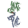
| ||||||||||||
| 単位格子 |
|
-要素
| #1: タンパク質 | 分子量: 47119.605 Da / 分子数: 2 / 由来タイプ: 組換発現 由来: (組換発現) 遺伝子: Irgb10 / 発現宿主:  #2: 化合物 |   分子量: 522.196 Da / 分子数: 2 / 由来タイプ: 合成 / 式: C10H17N6O13P3 / タイプ: SUBJECT OF INVESTIGATION 分子量: 522.196 Da / 分子数: 2 / 由来タイプ: 合成 / 式: C10H17N6O13P3 / タイプ: SUBJECT OF INVESTIGATIONコメント: GppNHp, GMPPNP, エネルギー貯蔵分子類似体*YM 研究の焦点であるリガンドがあるか | Y | Has protein modification | N | |
|---|
-実験情報
-実験
| 実験 | 手法: X線回折 / 使用した結晶の数: 1 |
|---|
- 試料調製
試料調製
| 結晶 | マシュー密度: 2.44 Å3/Da / 溶媒含有率: 49.59 % |
|---|---|
| 結晶化 | 温度: 293 K / 手法: 蒸気拡散法, ハンギングドロップ法 詳細: 20% (w/v) PEG 2,000 monomethyl ether (MME), 0.2 M trimethylamine n-oxide, 0.1 M Tris-HCl (pH 8.5) |
-データ収集
| 回折 | 平均測定温度: 100 K / Serial crystal experiment: N |
|---|---|
| 放射光源 | 由来: シンクロトロン / サイト: PAL/PLS / ビームライン: 5C (4A) / 波長: 1 Å |
| 検出器 | タイプ: DECTRIS EIGER2 X 9M / 検出器: PIXEL / 日付: 2022年11月29日 |
| 放射 | プロトコル: SINGLE WAVELENGTH / 単色(M)・ラウエ(L): M / 散乱光タイプ: x-ray |
| 放射波長 | 波長: 1 Å / 相対比: 1 |
| 反射 | 解像度: 3.05→29.43 Å / Num. obs: 17524 / % possible obs: 99.44 % / 冗長度: 6.6 % / Biso Wilson estimate: 84.19 Å2 / CC1/2: 0.998 / Net I/σ(I): 12.86 |
| 反射 シェル | 解像度: 3.05→3.159 Å / 冗長度: 6.8 % / Num. unique obs: 1743 / CC1/2: 0.866 / % possible all: 99.43 |
- 解析
解析
| ソフトウェア |
| |||||||||||||||||||||||||||||||||||||||||||||||||
|---|---|---|---|---|---|---|---|---|---|---|---|---|---|---|---|---|---|---|---|---|---|---|---|---|---|---|---|---|---|---|---|---|---|---|---|---|---|---|---|---|---|---|---|---|---|---|---|---|---|---|
| 精密化 | 構造決定の手法: 分子置換 / 解像度: 3.05→29.43 Å / SU ML: 0.5123 / 交差検証法: FREE R-VALUE / σ(F): 1.36 / 位相誤差: 31.6238 立体化学のターゲット値: GeoStd + Monomer Library + CDL v1.2
| |||||||||||||||||||||||||||||||||||||||||||||||||
| 溶媒の処理 | 減衰半径: 0.9 Å / VDWプローブ半径: 1.11 Å / 溶媒モデル: FLAT BULK SOLVENT MODEL | |||||||||||||||||||||||||||||||||||||||||||||||||
| 原子変位パラメータ | Biso mean: 93.03 Å2 | |||||||||||||||||||||||||||||||||||||||||||||||||
| 精密化ステップ | サイクル: LAST / 解像度: 3.05→29.43 Å
| |||||||||||||||||||||||||||||||||||||||||||||||||
| 拘束条件 |
| |||||||||||||||||||||||||||||||||||||||||||||||||
| LS精密化 シェル |
| |||||||||||||||||||||||||||||||||||||||||||||||||
| 精密化 TLS | 手法: refined / Origin x: 36.8027082296 Å / Origin y: -21.5252278279 Å / Origin z: 28.8807013495 Å
| |||||||||||||||||||||||||||||||||||||||||||||||||
| 精密化 TLSグループ | Selection details: all |
