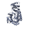
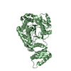
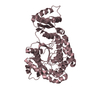
Entry Database : PDB / ID : 8jptTitle Crystal Structure of the acyltransferase domain from the eighth module of the spinosad polyketide synthase Polyene macrolide polyketide synthase Keywords / Function / homology Function Domain/homology Component
/ / / / / / / / / / / / / / / / / / / / / / / / / / / / / / / / / / / / / / / / / / / / / / / / / / / / / / / / / / / / / / / / / / / Biological species Saccharopolyspora spinosa (bacteria)Method / / / Resolution : 3.26 Å Authors Huang, S. / Zheng, J. Funding support Organization Grant number Country National Natural Science Foundation of China (NSFC) 32070040
Journal : Febs J. / Year : 2024Title : Structural and computational insights into the substrate specificity of acyltransferase domains from modular polyketide synthases.Authors : Huang, S. / Ji, H. / Zheng, J. History Deposition Jun 12, 2023 Deposition site / Processing site Revision 1.0 Jun 19, 2024 Provider / Type Revision 1.1 Jul 2, 2025 Group / Structure summary / Category / citation_author / pdbx_entry_detailsItem _citation.country / _citation.journal_abbrev ... _citation.country / _citation.journal_abbrev / _citation.journal_id_CSD / _citation.journal_id_ISSN / _citation.journal_volume / _citation.page_first / _citation.page_last / _citation.pdbx_database_id_DOI / _citation.pdbx_database_id_PubMed / _citation.title / _citation.year
Show all Show less
 Movie
Movie Controller

 Yorodumi
 Open data
 Basic information
 Components
 Keywords
 Function and homology information
 Saccharopolyspora spinosa (bacteria)
 X-RAY DIFFRACTION /
  Authors
 China, 1items
  Citation
 Structure visualization
 Downloads & links
 Other downloads
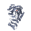
 PDBj
PDBj


 Assembly
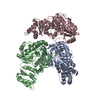
 Mass: 18.015 Da / Num. of mol.: 13 / Source method: isolated from a natural source / Formula: H2O
 Sample preparation
 Processing