 Movie
Movie Controller

[English] 日本語
 Yorodumi
- PDB-8jps: Structure of Duffy Antigen Receptor for Chemokines (DARC)/ACKR1 i... -
+ Open data

- Basic information
| Entry | Database: PDB / ID: 8jps | |||||||||||||||
|---|---|---|---|---|---|---|---|---|---|---|---|---|---|---|---|---|
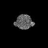
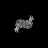
| Title | Structure of Duffy Antigen Receptor for Chemokines (DARC)/ACKR1 in complex with the chemokine, CCL7 (Composite map) | |||||||||||||||
 Components Components |
| |||||||||||||||
 Keywords Keywords | IMMUNE SYSTEM / GPCR / SIGNALING PROTEIN | |||||||||||||||
| Function / homology |  Function and homology information Function and homology informationregulation of chemokine production / CCR2 chemokine receptor binding / CCR1 chemokine receptor binding / positive regulation of natural killer cell chemotaxis / CCR chemokine receptor binding / chemokine-mediated signaling pathway / C-C chemokine binding / eosinophil chemotaxis / chemokine activity / Chemokine receptors bind chemokines ...regulation of chemokine production / CCR2 chemokine receptor binding / CCR1 chemokine receptor binding / positive regulation of natural killer cell chemotaxis / CCR chemokine receptor binding / chemokine-mediated signaling pathway / C-C chemokine binding / eosinophil chemotaxis / chemokine activity / Chemokine receptors bind chemokines / monocyte chemotaxis / cellular response to ethanol / cytoskeleton organization / Peptide ligand-binding receptors / response to gamma radiation / G protein-coupled receptor activity / defense response / recycling endosome / intracellular calcium ion homeostasis / chemotaxis / antimicrobial humoral immune response mediated by antimicrobial peptide / transmembrane signaling receptor activity / cell-cell signaling / regulation of cell shape / signaling receptor activity / heparin binding / early endosome / positive regulation of cell migration / inflammatory response / signal transduction / extracellular space / extracellular region / plasma membrane Similarity search - Function | |||||||||||||||
| Biological species |  Homo sapiens (human) Homo sapiens (human) | |||||||||||||||
| Method | ELECTRON MICROSCOPY / single particle reconstruction / cryo EM / Resolution: 3.65 Å | |||||||||||||||
 Authors Authors | Banerjee, R. / Khanppnavar, B. / Maharana, J. / Saha, S. / Korkhov, V.M. / Shukla, A.K. | |||||||||||||||
| Funding support |  India, India,  United Kingdom, 4items United Kingdom, 4items
| |||||||||||||||
 Citation Citation | Journal: Cell / Year: 2024 Title: Molecular mechanism of distinct chemokine engagement and functional divergence of the human Duffy antigen receptor. Authors: Shirsha Saha / Basavraj Khanppnavar / Jagannath Maharana / Heeryung Kim / Carlo Marion C Carino / Carole Daly / Shane Houston / Saloni Sharma / Nashrah Zaidi / Annu Dalal / Sudha Mishra / ...Authors: Shirsha Saha / Basavraj Khanppnavar / Jagannath Maharana / Heeryung Kim / Carlo Marion C Carino / Carole Daly / Shane Houston / Saloni Sharma / Nashrah Zaidi / Annu Dalal / Sudha Mishra / Manisankar Ganguly / Divyanshu Tiwari / Poonam Kumari / Gagan Deep Jhingan / Prem N Yadav / Bianca Plouffe / Asuka Inoue / Ka Young Chung / Ramanuj Banerjee / Volodymyr M Korkhov / Arun K Shukla /    Abstract: The Duffy antigen receptor is a seven-transmembrane (7TM) protein expressed primarily at the surface of red blood cells and displays strikingly promiscuous binding to multiple inflammatory and ...The Duffy antigen receptor is a seven-transmembrane (7TM) protein expressed primarily at the surface of red blood cells and displays strikingly promiscuous binding to multiple inflammatory and homeostatic chemokines. It serves as the basis of the Duffy blood group system in humans and also acts as the primary attachment site for malarial parasite Plasmodium vivax and pore-forming toxins secreted by Staphylococcus aureus. Here, we comprehensively profile transducer coupling of this receptor, discover potential non-canonical signaling pathways, and determine the cryoelectron microscopy (cryo-EM) structure in complex with the chemokine CCL7. The structure reveals a distinct binding mode of chemokines, as reflected by relatively superficial binding and a partially formed orthosteric binding pocket. We also observe a dramatic shortening of TM5 and 6 on the intracellular side, which precludes the formation of the docking site for canonical signal transducers, thereby providing a possible explanation for the distinct pharmacological and functional phenotype of this receptor. | |||||||||||||||
| History |
|
- Structure visualization
| Structure viewer | Molecule: MolmilJmol/JSmol |
|---|
- Downloads & links
-Download
| PDBx/mmCIF format | 8jps.cif.gz | 114.7 KB | Display | PDBx/mmCIF format |
|---|---|---|---|---|
| PDB format | pdb8jps.ent.gz | 86.4 KB | Display | PDB format |
| PDBx/mmJSON format | 8jps.json.gz | Tree view | PDBx/mmJSON format | |
| Others |  Other downloads Other downloads |
-Validation report
| Summary document | 8jps_validation.pdf.gz | 1.1 MB | Display | wwPDB validaton report |
|---|---|---|---|---|
| Full document | 8jps_full_validation.pdf.gz | 1.1 MB | Display | |
| Data in XML | 8jps_validation.xml.gz | 34.7 KB | Display | |
| Data in CIF | 8jps_validation.cif.gz | 49.3 KB | Display | |
| Arichive directory | https://data.pdbj.org/pub/pdb/validation_reports/jp/8jpsftp://data.pdbj.org/pub/pdb/validation_reports/jp/8jps | HTTPS FTP |
-Related structure data
| Related structure data | 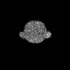 36488MC M: map data used to model this data C: citing same article ( |
|---|---|
| Similar structure data |
-Links
 PDBj
PDBj


- Assembly
| Deposited unit | 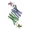
|
|---|---|
| 1 |
|
-Components
| #1: Protein | Mass: 28073.523 Da / Num. of mol.: 2 Source method: isolated from a genetically manipulated source Source: (gene. exp.) Homo sapiens (human) / Gene: ACKR1 / Production host:   Spodoptera frugiperda (fall armyworm) / References: UniProt: Q16570 Spodoptera frugiperda (fall armyworm) / References: UniProt: Q16570#2: Protein | Mass: 7594.902 Da / Num. of mol.: 2 Source method: isolated from a genetically manipulated source Source: (gene. exp.) Homo sapiens (human) / Gene: CCL7, MCP3, SCYA6, SCYA7 / Production host:  Has protein modification | Y | |
|---|
-Experimental details
-Experiment
| Experiment | Method: ELECTRON MICROSCOPY |
|---|---|
| EM experiment | Aggregation state: PARTICLE / 3D reconstruction method: single particle reconstruction |
- Sample preparation
| Component |
| ||||||||||||||||||||||||
|---|---|---|---|---|---|---|---|---|---|---|---|---|---|---|---|---|---|---|---|---|---|---|---|---|---|
| Molecular weight | Experimental value: NO | ||||||||||||||||||||||||
| Source (natural) | Organism: Homo sapiens (human) | ||||||||||||||||||||||||
| Source (recombinant) |
| ||||||||||||||||||||||||
| Buffer solution | pH: 7.4 | ||||||||||||||||||||||||
| Specimen | Embedding applied: NO / Shadowing applied: NO / Staining applied: NO / Vitrification applied: YES | ||||||||||||||||||||||||
| Vitrification | Cryogen name: ETHANE |
- Electron microscopy imaging
| Experimental equipment |  Model: Titan Krios / Image courtesy: FEI Company |
|---|---|
| Microscopy | Model: TFS KRIOS |
| Electron gun | Electron source:  FIELD EMISSION GUN / Accelerating voltage: 300 kV / Illumination mode: FLOOD BEAM FIELD EMISSION GUN / Accelerating voltage: 300 kV / Illumination mode: FLOOD BEAM |
| Electron lens | Mode: BRIGHT FIELD / Nominal defocus max: 2000 nm / Nominal defocus min: 1000 nm |
| Specimen holder | Cryogen: NITROGEN |
| Image recording | Electron dose: 58 e/Å2 / Film or detector model: GATAN K3 (6k x 4k) |
- Processing
| EM software |
| ||||||||||||||||||||||||
|---|---|---|---|---|---|---|---|---|---|---|---|---|---|---|---|---|---|---|---|---|---|---|---|---|---|
| CTF correction | Type: NONE | ||||||||||||||||||||||||
| Particle selection | Num. of particles selected: 25479169 | ||||||||||||||||||||||||
| Symmetry | Point symmetry: C2 (2 fold cyclic) | ||||||||||||||||||||||||
| 3D reconstruction | Resolution: 3.65 Å / Resolution method: FSC 0.143 CUT-OFF / Num. of particles: 308174 / Symmetry type: POINT | ||||||||||||||||||||||||
| Atomic model building | Protocol: FLEXIBLE FIT / Space: REAL | ||||||||||||||||||||||||
| Atomic model building | Source name: AlphaFold / Type: in silico model | ||||||||||||||||||||||||
| Refine LS restraints |
|
