National Natural Science Foundation of China (NSFC)
32271276
中国
引用
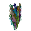
ジャーナル: Nat Commun / 年: 2023 タイトル: Serine peptidase Vpr forms enzymatically active fibrils outside Bacillus bacteria revealed by cryo-EM. 著者: Yijia Cheng / Jianting Han / Meinai Song / Shuqin Zhang / Qin Cao / 要旨: Bacteria develop a variety of extracellular fibrous structures crucial for their survival, such as flagella and pili. In this study, we use cryo-EM to identify protein fibrils surrounding lab- ...Bacteria develop a variety of extracellular fibrous structures crucial for their survival, such as flagella and pili. In this study, we use cryo-EM to identify protein fibrils surrounding lab-cultured Bacillus amyloiquefaciens and discover an unreported fibril species in addition to the flagellar fibrils. These previously unknown fibrils are composed of Vpr, an extracellular serine peptidase. We find that Vpr assembles into fibrils in an enzymatically active form, potentially representing a strategy of enriching Vpr activities around bacterial cells. Vpr fibrils are also observed under other culture conditions and around other Bacillus bacteria, such as Bacillus subtilis, which may suggest a general mechanism across all Bacillus bacterial groups. Taken together, our study reveals fibrils outside the bacterial cell and sheds light on the physiological role of these extracellular fibrils.
A: S8 family serine peptidase B: S8 family serine peptidase C: S8 family serine peptidase D: S8 family serine peptidase E: S8 family serine peptidase F: S8 family serine peptidase G: S8 family serine peptidase H: S8 family serine peptidase I: S8 family serine peptidase J: S8 family serine peptidase K: S8 family serine peptidase L: S8 family serine peptidase M: S8 family serine peptidase N: S8 family serine peptidase O: S8 family serine peptidase P: S8 family serine peptidase Q: S8 family serine peptidase R: S8 family serine peptidase
 ムービー
ムービー コントローラー


 データを開く
 基本情報
基本情報 要素
要素 キーワード
キーワード 機能・相同性情報
 データ登録者
 中国, 1件
  引用
引用 構造の表示
構造の表示 ダウンロードとリンク
 その他のダウンロード

 PDBj
PDBj


 集合体
集合体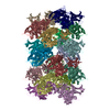
 試料調製
試料調製 電子顕微鏡撮影
 FIELD EMISSION GUN / 加速電圧: 300 kV / 照射モード: FLOOD BEAM
 解析
解析