 Movie
Movie Controller

[English] 日本語
 Yorodumi
- PDB-8jin: The local refined map of XBB spike protein (S) in complex with bi... -
+ Open data

- Basic information
| Entry | Database: PDB / ID: 8jin | ||||||
|---|---|---|---|---|---|---|---|
| Title | The local refined map of XBB spike protein (S) in complex with bispecific antibody G7-Fc | ||||||
 Components Components |
| ||||||
 Keywords Keywords | VIRAL PROTEIN / antibody | ||||||
| Function / homology |  Function and homology information Function and homology informationMaturation of spike protein / viral translation / Translation of Structural Proteins / Virion Assembly and Release / host cell surface / host extracellular space / suppression by virus of host tetherin activity / Induction of Cell-Cell Fusion / structural constituent of virion / entry receptor-mediated virion attachment to host cell ...Maturation of spike protein / viral translation / Translation of Structural Proteins / Virion Assembly and Release / host cell surface / host extracellular space / suppression by virus of host tetherin activity / Induction of Cell-Cell Fusion / structural constituent of virion / entry receptor-mediated virion attachment to host cell / host cell endoplasmic reticulum-Golgi intermediate compartment membrane / receptor-mediated endocytosis of virus by host cell / membrane fusion / Attachment and Entry / positive regulation of viral entry into host cell / receptor-mediated virion attachment to host cell / receptor ligand activity / host cell surface receptor binding / fusion of virus membrane with host plasma membrane / fusion of virus membrane with host endosome membrane / viral envelope / virion attachment to host cell / SARS-CoV-2 activates/modulates innate and adaptive immune responses / host cell plasma membrane / virion membrane / identical protein binding / membrane / plasma membrane Similarity search - Function | ||||||
| Biological species |  Homo sapiens (human) Homo sapiens (human)  Severe acute respiratory syndrome coronavirus 2 Severe acute respiratory syndrome coronavirus 2 | ||||||
| Method | ELECTRON MICROSCOPY / single particle reconstruction / cryo EM / Resolution: 2.45 Å | ||||||
 Authors Authors | Ma, Y. / Mao, Q. / Wang, Y. / Zhang, Z. | ||||||
| Funding support |  China, 1items China, 1items
| ||||||
 Citation Citation | Journal: To Be Published Title: Structure of XBB spike protein (S) di-trimer in complex with bispecific antibody G7-Fc at 3.75 Angstroms resolution. Authors: Ma, Y. / Mao, Q. / Wang, Y. / Zhang, Z. | ||||||
| History |
|
- Structure visualization
| Structure viewer | Molecule: MolmilJmol/JSmol |
|---|
- Downloads & links
-Download
| PDBx/mmCIF format | 8jin.cif.gz | 153.4 KB | Display | PDBx/mmCIF format |
|---|---|---|---|---|
| PDB format | pdb8jin.ent.gz | 104.5 KB | Display | PDB format |
| PDBx/mmJSON format | 8jin.json.gz | Tree view | PDBx/mmJSON format | |
| Others |  Other downloads Other downloads |
-Validation report
| Summary document | 8jin_validation.pdf.gz | 975 KB | Display | wwPDB validaton report |
|---|---|---|---|---|
| Full document | 8jin_full_validation.pdf.gz | 982.3 KB | Display | |
| Data in XML | 8jin_validation.xml.gz | 35.6 KB | Display | |
| Data in CIF | 8jin_validation.cif.gz | 49.4 KB | Display | |
| Arichive directory | https://data.pdbj.org/pub/pdb/validation_reports/ji/8jinftp://data.pdbj.org/pub/pdb/validation_reports/ji/8jin | HTTPS FTP |
-Related structure data
| Related structure data | 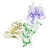 36321MC M: map data used to model this data C: citing same article ( |
|---|---|
| Similar structure data |
-Links
 PDBj
PDBj


- Assembly
| Deposited unit | 
|
|---|---|
| 1 |
|
-Components
| #1: Antibody | Mass: 26330.619 Da / Num. of mol.: 1 Source method: isolated from a genetically manipulated source Source: (gene. exp.) Homo sapiens (human) / Cell line (production host): HEK293F / Production host: Homo sapiens (human) |
|---|---|
| #2: Antibody | Mass: 27871.873 Da / Num. of mol.: 1 Source method: isolated from a genetically manipulated source Source: (gene. exp.) Homo sapiens (human) / Cell line (production host): HEK293F / Production host: Homo sapiens (human) |
| #3: Protein | Mass: 143147.531 Da / Num. of mol.: 1 Source method: isolated from a genetically manipulated source Source: (gene. exp.) Severe acute respiratory syndrome coronavirus 2Gene: S, 2 / Cell line (production host): HEK293F / Production host: Homo sapiens (human) / References: UniProt: P0DTC2 |
-Experimental details
-Experiment
| Experiment | Method: ELECTRON MICROSCOPY |
|---|---|
| EM experiment | Aggregation state: PARTICLE / 3D reconstruction method: single particle reconstruction |
- Sample preparation
| Component | Name: XBB spike protein (S) in complex with bispecific antibody G7-Fc Type: COMPLEX / Entity ID: all / Source: RECOMBINANT |
|---|---|
| Molecular weight | Experimental value: NO |
| Source (natural) | Organism: Severe acute respiratory syndrome coronavirus 2 |
| Source (recombinant) | Organism: Homo sapiens (human) |
| Details of virus | Isolate: STRAIN / Type: VIRION |
| Buffer solution | pH: 8 |
| Specimen | Embedding applied: NO / Shadowing applied: NO / Staining applied: NO / Vitrification applied: YES |
| Vitrification | Cryogen name: ETHANE |
- Electron microscopy imaging
| Experimental equipment |  Model: Titan Krios / Image courtesy: FEI Company |
|---|---|
| Microscopy | Model: FEI TITAN KRIOS |
| Electron gun | Electron source:  FIELD EMISSION GUN / Accelerating voltage: 300 kV / Illumination mode: FLOOD BEAM FIELD EMISSION GUN / Accelerating voltage: 300 kV / Illumination mode: FLOOD BEAM |
| Electron lens | Mode: BRIGHT FIELD / Nominal defocus max: 2200 nm / Nominal defocus min: 1200 nm |
| Image recording | Electron dose: 50 e/Å2 / Film or detector model: FEI FALCON IV (4k x 4k) |
- Processing
| CTF correction | Type: PHASE FLIPPING AND AMPLITUDE CORRECTION | ||||||||||||||||||||||||
|---|---|---|---|---|---|---|---|---|---|---|---|---|---|---|---|---|---|---|---|---|---|---|---|---|---|
| Symmetry | Point symmetry: C1 (asymmetric) | ||||||||||||||||||||||||
| 3D reconstruction | Resolution: 2.45 Å / Resolution method: FSC 0.143 CUT-OFF / Num. of particles: 838739 / Symmetry type: POINT | ||||||||||||||||||||||||
| Refine LS restraints |
|
