 Movie
Movie Controller


+ Open data

- Basic information
| Entry | Database: PDB / ID: 8jif | |||||||||||||||||||||||||||||||||||||||||||||
|---|---|---|---|---|---|---|---|---|---|---|---|---|---|---|---|---|---|---|---|---|---|---|---|---|---|---|---|---|---|---|---|---|---|---|---|---|---|---|---|---|---|---|---|---|---|---|
| Title | Cryo-EM Structure of 3-axis block of AAV9P31-Car4 complex | |||||||||||||||||||||||||||||||||||||||||||||
 Components Components |
| |||||||||||||||||||||||||||||||||||||||||||||
 Keywords Keywords | VIRAL PROTEIN/LYASE / receptor / aav9p31 / Carbonic anhydrases iv / Block-based reconstruction / VIRUS / VIRAL PROTEIN-LYASE complex | |||||||||||||||||||||||||||||||||||||||||||||
| Function / homology |  Function and homology information Function and homology informationErythrocytes take up carbon dioxide and release oxygen / Erythrocytes take up oxygen and release carbon dioxide / Reversible hydration of carbon dioxide / regulation of pH / bicarbonate transport / T=1 icosahedral viral capsid / transport vesicle membrane / endoplasmic reticulum-Golgi intermediate compartment / rough endoplasmic reticulum / secretory granule membrane ...Erythrocytes take up carbon dioxide and release oxygen / Erythrocytes take up oxygen and release carbon dioxide / Reversible hydration of carbon dioxide / regulation of pH / bicarbonate transport / T=1 icosahedral viral capsid / transport vesicle membrane / endoplasmic reticulum-Golgi intermediate compartment / rough endoplasmic reticulum / secretory granule membrane / carbonic anhydrase / carbonate dehydratase activity / sarcoplasmic reticulum / carbon dioxide transport / brush border membrane / trans-Golgi network / sarcolemma / apical plasma membrane / external side of plasma membrane / perinuclear region of cytoplasm / structural molecule activity / cell surface / Golgi apparatus / extracellular exosome / zinc ion binding / membrane / plasma membrane Similarity search - Function | |||||||||||||||||||||||||||||||||||||||||||||
| Biological species |    Adeno-associated virus 9 Adeno-associated virus 9 | |||||||||||||||||||||||||||||||||||||||||||||
| Method | ELECTRON MICROSCOPY / single particle reconstruction / cryo EM / Resolution: 2.28 Å | |||||||||||||||||||||||||||||||||||||||||||||
 Authors Authors | Zhang, R. / Liu, Y. / Lou, Z. | |||||||||||||||||||||||||||||||||||||||||||||
| Funding support |  China, 3items China, 3items
| |||||||||||||||||||||||||||||||||||||||||||||
 Citation Citation | Journal: PLoS Pathog / Year: 2024 Title: Structural basis of the recognition of adeno-associated virus by the neurological system-related receptor carbonic anhydrase IV. Authors: Ran Zhang / Yixiao Liu / Fengxi Yu / Guangxue Xu / Lili Li / Baobin Li / Zhiyong Lou /  Abstract: Carbonic anhydrase IV (Car4) is a newly identified receptor that allows adeno-associated virus (AAV) 9P31 to cross the blood-brain barrier and achieve efficient infection in the central nervous ...Carbonic anhydrase IV (Car4) is a newly identified receptor that allows adeno-associated virus (AAV) 9P31 to cross the blood-brain barrier and achieve efficient infection in the central nervous system (CNS) in mouse models. However, the molecular mechanism by which engineered AAV capsids with 7-mer insertion in the variable region (VR) VIII recognize these novel cellular receptors is unknown. Here we report the cryo-EM structures of AAV9P31 and its complex with Mus musculus Car4 at atomic resolution by utilizing the block-based reconstruction (BBR) method. The structures demonstrated that Car4 binds to the protrusions at 3-fold axes of the capsid. The inserted 7-mer extends into a hydrophobic region near the catalytic center of Car4 to form stable interactions. Mutagenesis studies also identified the key residues in Car4 responsible for the AAV9P31 interaction. These findings provide new insights into the novel receptor recognition mechanism of AAV generated by directed evolution and highlight the application of the BBR method to studying the virus-receptor molecular mechanism. | |||||||||||||||||||||||||||||||||||||||||||||
| History |
|
- Structure visualization
| Structure viewer | Molecule: MolmilJmol/JSmol |
|---|
- Downloads & links
-Download
| PDBx/mmCIF format | 8jif.cif.gz | 335.5 KB | Display | PDBx/mmCIF format |
|---|---|---|---|---|
| PDB format | pdb8jif.ent.gz | Display | PDB format | |
| PDBx/mmJSON format | 8jif.json.gz | Tree view | PDBx/mmJSON format | |
| Others |  Other downloads Other downloads |
-Validation report
| Arichive directory | https://data.pdbj.org/pub/pdb/validation_reports/ji/8jifftp://data.pdbj.org/pub/pdb/validation_reports/ji/8jif | HTTPS FTP |
|---|
-Related structure data
| Related structure data |  36311MC  8xegC M: map data used to model this data C: citing same article ( |
|---|---|
| Similar structure data |

-Links
 PDBj
PDBj


- Assembly
| Deposited unit | 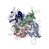
|
|---|---|
| 1 |
|
-Components
| #1: Protein | Mass: 29281.367 Da / Num. of mol.: 1 Source method: isolated from a genetically manipulated source Source: (gene. exp.) Production host:  References: UniProt: Q64444, carbonic anhydrase | ||||||
|---|---|---|---|---|---|---|---|
| #2: Protein | Mass: 59295.277 Da / Num. of mol.: 3 Source method: isolated from a genetically manipulated source Source: (gene. exp.) Adeno-associated virus 9 / Gene: cap / Production host:  Homo sapiens (human) / References: UniProt: Q6JC40 Homo sapiens (human) / References: UniProt: Q6JC40#3: Chemical | ChemComp-ZN / |   Mass: 65.409 Da / Num. of mol.: 1 / Source method: obtained synthetically / Formula: Zn / Feature type: SUBJECT OF INVESTIGATION Mass: 65.409 Da / Num. of mol.: 1 / Source method: obtained synthetically / Formula: Zn / Feature type: SUBJECT OF INVESTIGATIONHas ligand of interest | Y | Has protein modification | Y | |
-Experimental details
-Experiment
| Experiment | Method: ELECTRON MICROSCOPY |
|---|---|
| EM experiment | Aggregation state: PARTICLE / 3D reconstruction method: single particle reconstruction |
- Sample preparation
| Component | Name: 3-fold axes block of AAV9P31 and Car4 complex aat 2.28 angstrom Type: COMPLEX / Entity ID: #1-#2 / Source: RECOMBINANT | |||||||||||||||
|---|---|---|---|---|---|---|---|---|---|---|---|---|---|---|---|---|
| Molecular weight | Experimental value: NO | |||||||||||||||
| Source (natural) |
| |||||||||||||||
| Source (recombinant) |
| |||||||||||||||
| Buffer solution | pH: 8 / Details: 20mM Tris-Cl, pH8.0; 150mM NaCl. | |||||||||||||||
| Buffer component |
| |||||||||||||||
| Specimen | Conc.: 2 mg/ml / Embedding applied: NO / Shadowing applied: NO / Staining applied: NO / Vitrification applied: YES | |||||||||||||||
| Specimen support | Grid material: COPPER / Grid mesh size: 300 divisions/in. / Grid type: Quantifoil R0.6/1 | |||||||||||||||
| Vitrification | Instrument: FEI VITROBOT MARK III / Cryogen name: ETHANE / Humidity: 100 % / Chamber temperature: 280 K |
- Electron microscopy imaging
| Experimental equipment |  Model: Titan Krios / Image courtesy: FEI Company |
|---|---|
| Microscopy | Model: FEI TITAN KRIOS |
| Electron gun | Electron source:  FIELD EMISSION GUN / Accelerating voltage: 300 kV / Illumination mode: FLOOD BEAM FIELD EMISSION GUN / Accelerating voltage: 300 kV / Illumination mode: FLOOD BEAM |
| Electron lens | Mode: BRIGHT FIELD / Nominal defocus max: 1800 nm / Nominal defocus min: 600 nm |
| Image recording | Electron dose: 50 e/Å2 / Film or detector model: GATAN K2 QUANTUM (4k x 4k) |
- Processing
| EM software |
| ||||||||||||||||||||||||
|---|---|---|---|---|---|---|---|---|---|---|---|---|---|---|---|---|---|---|---|---|---|---|---|---|---|
| CTF correction | Type: NONE | ||||||||||||||||||||||||
| 3D reconstruction | Resolution: 2.28 Å / Resolution method: FSC 0.143 CUT-OFF / Num. of particles: 13604676 / Symmetry type: POINT | ||||||||||||||||||||||||
| Refine LS restraints |
|
