[English] 日本語
Yorodumi
- PDB-8jg7: Serine decarboxylase -

+
Open data


ID or keywords:

Loading...

-
Basic information

Entry
Database: PDB / ID: 8jg7
TitleSerine decarboxylase
ComponentsSerine decarboxylase
KeywordsBIOSYNTHETIC PROTEIN / PLP
Function / homology
Function and homology information


ethanolamine metabolic process / serine decarboxylase activity / Lyases; Carbon-carbon lyases; Carboxy-lyases / carboxylic acid metabolic process / pyridoxal phosphate binding / plasma membrane / cytosol
Similarity search - Function
Pyridoxal-phosphate binding site / DDC / GAD / HDC / TyrDC pyridoxal-phosphate attachment site. / Pyridoxal phosphate-dependent decarboxylase / Pyridoxal-dependent decarboxylase conserved domain / Pyridoxal phosphate-dependent transferase, small domain / Pyridoxal phosphate-dependent transferase, major domain / Pyridoxal phosphate-dependent transferase
Similarity search - Domain/homology
PYRIDOXAL-5'-PHOSPHATE / Serine decarboxylase
Similarity search - Component
Biological speciesArabidopsis thaliana (thale cress)
MethodX-RAY DIFFRACTION / SYNCHROTRON / MOLECULAR REPLACEMENT / Resolution: 2.85 Å
AuthorsWang, H. / Gong, W.
Funding support1items
OrganizationGrant numberCountry
Other government
CitationJournal: To Be Published
Title: Crystal structure of aserine decarboxylase from Arabidopsis thaliana
Authors: Wang, H.
History
DepositionMay 19, 2023Deposition site: PDBJ / Processing site: PDBJ
Revision 1.0May 22, 2024Provider: repository / Type: Initial release

-
Structure visualization

Structure viewerMolecule:
MolmilJmol/JSmol

Downloads & links

-
Assembly

Deposited unit
A: Serine decarboxylase
C: Serine decarboxylase
D: Serine decarboxylase
E: Serine decarboxylase
hetero molecules


Theoretical massNumber of molelcules
Total (without water)191,36416
Polymers189,7454
Non-polymers1,61912
Water00
1
A: Serine decarboxylase
E: Serine decarboxylase
hetero molecules


Theoretical massNumber of molelcules
Total (without water)95,6828
Polymers94,8722
Non-polymers8096
Water0
TypeNameSymmetry operationNumber
identity operation1_555x,y,z1
Buried area9870 Å2
ΔGint-53 kcal/mol
Surface area31050 Å2
MethodPISA
2
C: Serine decarboxylase
D: Serine decarboxylase
hetero molecules


Theoretical massNumber of molelcules
Total (without water)95,6828
Polymers94,8722
Non-polymers8096
Water0
TypeNameSymmetry operationNumber
identity operation1_555x,y,z1
Buried area9850 Å2
ΔGint-54 kcal/mol
Surface area31120 Å2
MethodPISA
Unit cell
Length a, b, c (Å)115.360, 149.450, 139.126
Angle α, β, γ (deg.)90.00, 109.18, 90.00
Int Tables number5
Space group name H-MC121

-
Components

#1: Protein
Serine decarboxylase / AtSDC / Protein EMBRYO DEFECTIVE 1075 / Serine decarboxylase 1 / AtSDC1


Mass: 47436.230 Da / Num. of mol.: 4
Source method: isolated from a genetically manipulated source
Source: (gene. exp.) Arabidopsis thaliana (thale cress) / Gene: SDC, EMB1075, SDC1, At1g43710, F2J6.7 / Production host: Escherichia coli (E. coli)
References: UniProt: Q9MA74, Lyases; Carbon-carbon lyases; Carboxy-lyases
#2: Chemical
ChemComp-PLP / PYRIDOXAL-5'-PHOSPHATE / VITAMIN B6 Phosphate


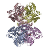
Mass: 247.142 Da / Num. of mol.: 4 / Source method: obtained synthetically / Formula: C8H10NO6P / Feature type: SUBJECT OF INVESTIGATION
#3: Chemical
ChemComp-ZN / ZINC ION


Mass: 65.409 Da / Num. of mol.: 4 / Source method: obtained synthetically / Formula: Zn
#4: Chemical
ChemComp-GOL / GLYCEROL / GLYCERIN / PROPANE-1,2,3-TRIOL


Mass: 92.094 Da / Num. of mol.: 4 / Source method: obtained synthetically / Formula: C3H8O3
Has ligand of interestY

-
Experimental details

-
Experiment

ExperimentMethod: X-RAY DIFFRACTION / Number of used crystals: 1

-
Sample preparation

CrystalDensity Matthews: 2.99 Å3/Da / Density % sol: 58.85 %
Crystal growTemperature: 289.15 K / Method: vapor diffusion, sitting drop / Details: 20% PEG 400, 0.2M CaCl2, PH8.0

-
Data collection

DiffractionMean temperature: 100 K / Serial crystal experiment: N
Diffraction sourceSource: SYNCHROTRON / Site: SSRF / Beamline: BL18U1 / Wavelength: 0.979 Å
DetectorType: DECTRIS PILATUS3 6M / Detector: PIXEL / Date: May 16, 2021
RadiationProtocol: SINGLE WAVELENGTH / Monochromatic (M) / Laue (L): M / Scattering type: x-ray
Radiation wavelengthWavelength: 0.979 Å / Relative weight: 1
ReflectionResolution: 2.85→50 Å / Num. obs: 52125 / % possible obs: 99.6 % / Redundancy: 5.9 % / CC1/2: 0.86 / CC star: 0.962 / Rmerge(I) obs: 0.173 / Rpim(I) all: 0.076 / Rrim(I) all: 0.189 / Χ2: 2.023 / Net I/σ(I): 3.5
Reflection shell

Diffraction-ID: 1

Resolution (Å)Redundancy (%)Rmerge(I) obsNum. unique obsCC1/2CC starRpim(I) allRrim(I) allΧ2% possible all
2.85-2.950.55125560.8460.9570.270.6160.54598.1
2.9-2.955.70.54926170.8980.9730.2520.6060.55699.9
2.95-3.016.20.49625800.9160.9780.2160.5410.561100
3.01-3.076.10.42125950.9190.9790.1840.460.5999.9
3.07-3.146.20.36526230.9450.9860.1580.3990.732100
3.14-3.216.10.32526050.9520.9880.1410.3550.72899.9
3.21-3.296.10.28625810.9660.9910.1240.3130.70799.9
3.29-3.386.10.25826330.9640.9910.1120.2820.813100
3.38-3.486.10.2826100.3540.7230.1280.3091.118100
3.48-3.596.10.18525870.9770.9940.080.2021.04599.9
3.59-3.726.10.1826240.9810.9950.0790.1971.141100
3.72-3.8760.16426000.9690.9920.0710.1791.50899.9
3.87-4.0460.16225830.9780.9940.0710.1771.5199.7
4.04-4.265.90.13326250.9720.9930.0580.1461.79399.6
4.26-4.525.80.1326210.9640.9910.0580.1431.90999.5
4.52-4.875.70.11625790.9810.9950.0520.1271.80599.7
4.87-5.365.20.11926070.9720.9930.0550.1312.52399.1
5.36-6.145.50.12825700.9610.990.0570.1412.89498
6.14-7.736.50.11226440.9720.9930.0470.1213.101100
7.73-506.40.16526850.9040.9750.070.1813.552100

-
Processing

Software
NameVersionClassification
REFMAC5.8.0352refinement
HKL-3000data reduction
HKL-3000data scaling
PHASERphasing
RefinementMethod to determine structure: MOLECULAR REPLACEMENT / Resolution: 2.85→49.39 Å / Cor.coef. Fo:Fc: 0.922 / Cor.coef. Fo:Fc free: 0.886 / SU B: 16.235 / SU ML: 0.304 / Cross valid method: THROUGHOUT / ESU R Free: 0.39 / Stereochemistry target values: MAXIMUM LIKELIHOOD / Details: HYDROGENS HAVE BEEN ADDED IN THE RIDING POSITIONS
RfactorNum. reflection% reflectionSelection details
Rfree0.2618 2576 4.9 %RANDOM
Rwork0.21353 ---
obs0.21591 49543 99.42 %-
Solvent computationIon probe radii: 0.8 Å / Shrinkage radii: 0.8 Å / VDW probe radii: 1.2 Å / Solvent model: MASK
Displacement parametersBiso mean: 55.93 Å2
Baniso -1Baniso -2Baniso -3
1-7.32 Å2-0 Å2-0.2 Å2
2---1.58 Å20 Å2
3----4.51 Å2
Refinement stepCycle: 1 / Resolution: 2.85→49.39 Å
ProteinNucleic acidLigandSolventTotal
Num. atoms13000 0 88 0 13088
Refine LS restraints
Refine-IDTypeDev idealDev ideal targetNumber
X-RAY DIFFRACTIONr_bond_refined_d0.0070.01213404
X-RAY DIFFRACTIONr_bond_other_d0.0020.01612142
X-RAY DIFFRACTIONr_angle_refined_deg1.411.6418108
X-RAY DIFFRACTIONr_angle_other_deg0.4651.55828291
X-RAY DIFFRACTIONr_dihedral_angle_1_deg7.31751624
X-RAY DIFFRACTIONr_dihedral_angle_2_deg15.8311092
X-RAY DIFFRACTIONr_dihedral_angle_3_deg18.36102304
X-RAY DIFFRACTIONr_dihedral_angle_4_deg
X-RAY DIFFRACTIONr_chiral_restr0.0660.21944
X-RAY DIFFRACTIONr_gen_planes_refined0.0060.0215204
X-RAY DIFFRACTIONr_gen_planes_other0.0010.022728
X-RAY DIFFRACTIONr_nbd_refined
X-RAY DIFFRACTIONr_nbd_other
X-RAY DIFFRACTIONr_nbtor_refined
X-RAY DIFFRACTIONr_nbtor_other
X-RAY DIFFRACTIONr_xyhbond_nbd_refined
X-RAY DIFFRACTIONr_xyhbond_nbd_other
X-RAY DIFFRACTIONr_metal_ion_refined
X-RAY DIFFRACTIONr_metal_ion_other
X-RAY DIFFRACTIONr_symmetry_vdw_refined
X-RAY DIFFRACTIONr_symmetry_vdw_other
X-RAY DIFFRACTIONr_symmetry_hbond_refined
X-RAY DIFFRACTIONr_symmetry_hbond_other
X-RAY DIFFRACTIONr_symmetry_metal_ion_refined
X-RAY DIFFRACTIONr_symmetry_metal_ion_other
X-RAY DIFFRACTIONr_mcbond_it5.0645.5186520
X-RAY DIFFRACTIONr_mcbond_other5.0615.5186520
X-RAY DIFFRACTIONr_mcangle_it7.5668.2528136
X-RAY DIFFRACTIONr_mcangle_other7.5658.2538137
X-RAY DIFFRACTIONr_scbond_it5.3775.9236884
X-RAY DIFFRACTIONr_scbond_other5.3755.9246881
X-RAY DIFFRACTIONr_scangle_it
X-RAY DIFFRACTIONr_scangle_other8.0888.7239973
X-RAY DIFFRACTIONr_long_range_B_refined10.44869.42814924
X-RAY DIFFRACTIONr_long_range_B_other10.44869.42814925
X-RAY DIFFRACTIONr_rigid_bond_restr
X-RAY DIFFRACTIONr_sphericity_free
X-RAY DIFFRACTIONr_sphericity_bonded
LS refinement shellResolution: 2.85→2.916 Å
RfactorNum. reflection% reflection
Rfree0.331 177 -
Rwork0.294 3527 -
obs--95.98 %

+
About Yorodumi

-
News

-
Feb 9, 2022. New format data for meta-information of EMDB entries

New format data for meta-information of EMDB entries

  • Version 3 of the EMDB header file is now the official format.
  • The previous official version 1.9 will be removed from the archive.

Related info.:EMDB header

External links:wwPDB to switch to version 3 of the EMDB data model

-
Aug 12, 2020. Covid-19 info

Covid-19 info

URL: https://pdbjlvh1.pdbj.org/emnavi/covid19.php

New page: Covid-19 featured information page in EM Navigator.

Related info.:Covid-19 info / Mar 5, 2020. Novel coronavirus structure data

+
Mar 5, 2020. Novel coronavirus structure data

Novel coronavirus structure data

Related info.:Yorodumi Speices / Aug 12, 2020. Covid-19 info

External links:COVID-19 featured content - PDBj / Molecule of the Month (242):Coronavirus Proteases

+
Jan 31, 2019. EMDB accession codes are about to change! (news from PDBe EMDB page)

EMDB accession codes are about to change! (news from PDBe EMDB page)

  • The allocation of 4 digits for EMDB accession codes will soon come to an end. Whilst these codes will remain in use, new EMDB accession codes will include an additional digit and will expand incrementally as the available range of codes is exhausted. The current 4-digit format prefixed with “EMD-” (i.e. EMD-XXXX) will advance to a 5-digit format (i.e. EMD-XXXXX), and so on. It is currently estimated that the 4-digit codes will be depleted around Spring 2019, at which point the 5-digit format will come into force.
  • The EM Navigator/Yorodumi systems omit the EMD- prefix.

Related info.:Q: What is EMD? / ID/Accession-code notation in Yorodumi/EM Navigator

External links:EMDB Accession Codes are Changing Soon! / Contact to PDBj

+
Jul 12, 2017. Major update of PDB

Major update of PDB

  • wwPDB released updated PDB data conforming to the new PDBx/mmCIF dictionary.
  • This is a major update changing the version number from 4 to 5, and with Remediation, in which all the entries are updated.
  • In this update, many items about electron microscopy experimental information are reorganized (e.g. em_software).
  • Now, EM Navigator and Yorodumi are based on the updated data.

External links:wwPDB Remediation / Enriched Model Files Conforming to OneDep Data Standards Now Available in the PDB FTP Archive

-
Yorodumi

Thousand views of thousand structures

  • Yorodumi is a browser for structure data from EMDB, PDB, SASBDB, etc.
  • This page is also the successor to EM Navigator detail page, and also detail information page/front-end page for Omokage search.
  • The word "yorodu" (or yorozu) is an old Japanese word meaning "ten thousand". "mi" (miru) is to see.

Related info.:EMDB / PDB / SASBDB / Comparison of 3 databanks / Yorodumi Search / Aug 31, 2016. New EM Navigator & Yorodumi / Yorodumi Papers / Jmol/JSmol / Function and homology information / Changes in new EM Navigator and Yorodumi

Read more