 Movie
Movie Controller


+ Open data

- Basic information
| Entry | Database: PDB / ID: 8jc1 | ||||||
|---|---|---|---|---|---|---|---|
| Title | Crystal structure of Pectocin M1 from Pectobacterium carotovorum | ||||||
 Components Components | pectocin M1 | ||||||
 Keywords Keywords | HYDROLASE / bactereocin / pectocin M1 / colicin / ferredoxin-like protein / peptidoglycan | ||||||
| Function / homology |  Function and homology information Function and homology informationelectron transport chain / 2 iron, 2 sulfur cluster binding / electron transfer activity / defense response to bacterium / metal ion binding Similarity search - Function | ||||||
| Biological species |  Pectobacterium carotovorum (bacteria) Pectobacterium carotovorum (bacteria) | ||||||
| Method |  X-RAY DIFFRACTION / SYNCHROTRON / MOLECULAR REPLACEMENT / Resolution: 2.04 Å X-RAY DIFFRACTION / SYNCHROTRON / MOLECULAR REPLACEMENT / Resolution: 2.04 Å | ||||||
 Authors Authors | Jantarit, N. / Kurisu, G. / Tanaka, H. | ||||||
| Funding support |  Japan, 1items Japan, 1items
| ||||||
 Citation Citation | Journal: Febs Open Bio / Year: 2024 Title: Crystal structure of pectocin M1 reveals diverse conformations and interactions during its initial step via the ferredoxin uptake system. Authors: Jantarit, N. / Tanaka, H. / Lin, Y. / Lee, Y.H. / Kurisu, G. | ||||||
| History |
|
- Structure visualization
| Structure viewer | Molecule: MolmilJmol/JSmol |
|---|
- Downloads & links
-Download
| PDBx/mmCIF format | 8jc1.cif.gz | 278.4 KB | Display | PDBx/mmCIF format |
|---|---|---|---|---|
| PDB format | pdb8jc1.ent.gz | 180.6 KB | Display | PDB format |
| PDBx/mmJSON format | 8jc1.json.gz | Tree view | PDBx/mmJSON format | |
| Others |  Other downloads Other downloads |
-Validation report
| Summary document | 8jc1_validation.pdf.gz | 3.5 MB | Display | wwPDB validaton report |
|---|---|---|---|---|
| Full document | 8jc1_full_validation.pdf.gz | 3.5 MB | Display | |
| Data in XML | 8jc1_validation.xml.gz | 51.9 KB | Display | |
| Data in CIF | 8jc1_validation.cif.gz | 69.6 KB | Display | |
| Arichive directory | https://data.pdbj.org/pub/pdb/validation_reports/jc/8jc1ftp://data.pdbj.org/pub/pdb/validation_reports/jc/8jc1 | HTTPS FTP |
-Related structure data
| Related structure data | 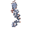 4n58S S: Starting model for refinement |
|---|---|
| Similar structure data |
-Links
 PDBj
PDBj


- Assembly
| Deposited unit | 
| ||||||||||||
|---|---|---|---|---|---|---|---|---|---|---|---|---|---|
| 1 |
| ||||||||||||
| Unit cell |
|
-Components
-Protein , 1 types, 4 molecules ABCD
| #1: Protein | Mass: 29354.586 Da / Num. of mol.: 4 Source method: isolated from a genetically manipulated source Source: (gene. exp.) Pectobacterium carotovorum (bacteria) / Strain: PC1 / Gene: PC1_2303 / Plasmid: pET28a / Production host: |
|---|
-Non-polymers , 8 types, 565 molecules 


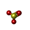


| #2: Chemical | ChemComp-FES /  Mass: 175.820 Da / Num. of mol.: 4 / Source method: obtained synthetically / Formula: Fe2S2 / Feature type: SUBJECT OF INVESTIGATION Mass: 175.820 Da / Num. of mol.: 4 / Source method: obtained synthetically / Formula: Fe2S2 / Feature type: SUBJECT OF INVESTIGATION#3: Chemical | ChemComp-NA /  Mass: 22.990 Da / Num. of mol.: 8 / Source method: obtained synthetically / Formula: Na Mass: 22.990 Da / Num. of mol.: 8 / Source method: obtained synthetically / Formula: Na#4: Chemical | ChemComp-MG /  Mass: 24.305 Da / Num. of mol.: 4 / Source method: obtained synthetically / Formula: Mg / Feature type: SUBJECT OF INVESTIGATION Mass: 24.305 Da / Num. of mol.: 4 / Source method: obtained synthetically / Formula: Mg / Feature type: SUBJECT OF INVESTIGATION#5: Chemical |  Mass: 80.063 Da / Num. of mol.: 2 / Source method: obtained synthetically / Formula: SO3 Mass: 80.063 Da / Num. of mol.: 2 / Source method: obtained synthetically / Formula: SO3#6: Chemical | ChemComp-GOL /  Mass: 92.094 Da / Num. of mol.: 4 / Source method: obtained synthetically / Formula: C3H8O3 Mass: 92.094 Da / Num. of mol.: 4 / Source method: obtained synthetically / Formula: C3H8O3#7: Chemical | ChemComp-SO4 / |  Mass: 96.063 Da / Num. of mol.: 1 / Source method: obtained synthetically / Formula: SO4 Mass: 96.063 Da / Num. of mol.: 1 / Source method: obtained synthetically / Formula: SO4#8: Chemical |  Mass: 35.453 Da / Num. of mol.: 2 / Source method: obtained synthetically / Formula: Cl Mass: 35.453 Da / Num. of mol.: 2 / Source method: obtained synthetically / Formula: Cl#9: Water | ChemComp-HOH / | Mass: 18.015 Da / Num. of mol.: 540 / Source method: isolated from a natural source / Formula: H2O |
|---|
-Details
| Has ligand of interest | Y |
|---|---|
| Has protein modification | N |
-Experimental details
-Experiment
| Experiment | Method: X-RAY DIFFRACTION / Number of used crystals: 1 |
|---|
- Sample preparation
| Crystal | Density Matthews: 2.69 Å3/Da / Density % sol: 54.26 % |
|---|---|
| Crystal grow | Temperature: 293.15 K / Method: vapor diffusion, hanging drop / pH: 7.5 Details: 0.1 M HEPES pH 7.5, 19.2% w/v poly(acrylic acid sodium salt) 5,100 |
-Data collection
| Diffraction | Mean temperature: 100 K / Serial crystal experiment: N |
|---|---|
| Diffraction source | Source: SYNCHROTRON / Site: SPring-8 / Beamline: BL44XU / Wavelength: 0.9 Å |
| Detector | Type: DECTRIS EIGER X 16M / Detector: PIXEL / Date: Apr 12, 2023 |
| Radiation | Protocol: SINGLE WAVELENGTH / Monochromatic (M) / Laue (L): M / Scattering type: x-ray |
| Radiation wavelength | Wavelength: 0.9 Å / Relative weight: 1 |
| Reflection | Resolution: 2.04→49.49 Å / Num. obs: 77569 / % possible obs: 95.5 % / Redundancy: 1.82 % / Biso Wilson estimate: 38.86 Å2 / CC1/2: 0.993 / CC star: 0 / Rmerge(I) obs: 0.077 / Net I/σ(I): 6.63 |
| Reflection shell | Resolution: 2.04→2.17 Å / Rmerge(I) obs: 0.53 / Mean I/σ(I) obs: 1.17 / Num. unique obs: 23492 / CC1/2: 0.7 / % possible all: 87.8 |
- Processing
| Software |
| |||||||||||||||||||||||||||||||||||||||||||||||||||||||||||||||||||||||||||||||||||||||||||||||||||||||||
|---|---|---|---|---|---|---|---|---|---|---|---|---|---|---|---|---|---|---|---|---|---|---|---|---|---|---|---|---|---|---|---|---|---|---|---|---|---|---|---|---|---|---|---|---|---|---|---|---|---|---|---|---|---|---|---|---|---|---|---|---|---|---|---|---|---|---|---|---|---|---|---|---|---|---|---|---|---|---|---|---|---|---|---|---|---|---|---|---|---|---|---|---|---|---|---|---|---|---|---|---|---|---|---|---|---|---|
| Refinement | Method to determine structure: MOLECULAR REPLACEMENT Starting model: 4N58 Resolution: 2.04→49.49 Å / SU ML: 0.2753 / Cross valid method: FREE R-VALUE / σ(F): 1.36 / Phase error: 25.7312 Stereochemistry target values: GeoStd + Monomer Library + CDL v1.2
| |||||||||||||||||||||||||||||||||||||||||||||||||||||||||||||||||||||||||||||||||||||||||||||||||||||||||
| Solvent computation | Shrinkage radii: 0.9 Å / VDW probe radii: 1.11 Å / Solvent model: FLAT BULK SOLVENT MODEL | |||||||||||||||||||||||||||||||||||||||||||||||||||||||||||||||||||||||||||||||||||||||||||||||||||||||||
| Displacement parameters | Biso mean: 46.94 Å2 | |||||||||||||||||||||||||||||||||||||||||||||||||||||||||||||||||||||||||||||||||||||||||||||||||||||||||
| Refinement step | Cycle: LAST / Resolution: 2.04→49.49 Å
| |||||||||||||||||||||||||||||||||||||||||||||||||||||||||||||||||||||||||||||||||||||||||||||||||||||||||
| Refine LS restraints |
| |||||||||||||||||||||||||||||||||||||||||||||||||||||||||||||||||||||||||||||||||||||||||||||||||||||||||
| LS refinement shell |
|
