[English] 日本語
Yorodumi
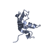
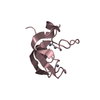
- PDB-8j9q: Crystal structure of UBR box of UBR4 apo -

+
Open data


ID or keywords:

Loading...

-
Basic information

Entry
Database: PDB / ID: 8j9q
TitleCrystal structure of UBR box of UBR4 apo
ComponentsE3 ubiquitin-protein ligase UBR4
KeywordsLIGASE / E3 ligase / UBR box
Function / homology
Function and homology information


negative regulation of fatty acid biosynthetic process / tertiary granule membrane / ficolin-1-rich granule membrane / specific granule membrane / RING-type E3 ubiquitin transferase / ubiquitin-protein transferase activity / ubiquitin protein ligase activity / Antigen processing: Ubiquitination & Proteasome degradation / ubiquitin-dependent protein catabolic process / protein ubiquitination ...negative regulation of fatty acid biosynthetic process / tertiary granule membrane / ficolin-1-rich granule membrane / specific granule membrane / RING-type E3 ubiquitin transferase / ubiquitin-protein transferase activity / ubiquitin protein ligase activity / Antigen processing: Ubiquitination & Proteasome degradation / ubiquitin-dependent protein catabolic process / protein ubiquitination / calmodulin binding / centrosome / Neutrophil degranulation / zinc ion binding / nucleoplasm / membrane / plasma membrane / cytosol
Similarity search - Function
E3 ubiquitin-protein ligase UBR4, N-terminal / : / E3 ubiquitin-protein ligase UBR4 N-terminal / E3 ubiquitin ligase UBR4, C-terminal / E3 ubiquitin ligase UBR4-like / E3 ubiquitin-protein ligase UBR4 / Putative zinc finger in N-recognin (UBR box) / Zinc finger, UBR-type / Zinc finger UBR-type profile. / Putative zinc finger in N-recognin, a recognition component of the N-end rule pathway ...E3 ubiquitin-protein ligase UBR4, N-terminal / : / E3 ubiquitin-protein ligase UBR4 N-terminal / E3 ubiquitin ligase UBR4, C-terminal / E3 ubiquitin ligase UBR4-like / E3 ubiquitin-protein ligase UBR4 / Putative zinc finger in N-recognin (UBR box) / Zinc finger, UBR-type / Zinc finger UBR-type profile. / Putative zinc finger in N-recognin, a recognition component of the N-end rule pathway / Armadillo-type fold / WD40-repeat-containing domain superfamily
Similarity search - Domain/homology
E3 ubiquitin-protein ligase UBR4
Similarity search - Component
Biological speciesHomo sapiens (human)
MethodX-RAY DIFFRACTION / SYNCHROTRON / MOLECULAR REPLACEMENT / Resolution: 2.18 Å
AuthorsJeong, D.-E. / KIm, S.-J. / Shin, H.-C.
Funding support Korea, Republic Of, 3items
OrganizationGrant numberCountry
Other governmentCRC22021-700 Korea, Republic Of
Other governmentKGM9952314 Korea, Republic Of
Other government2021M3A9G8025599 Korea, Republic Of
CitationJournal: Commun Biol / Year: 2023
Title: Insights into the recognition mechanism in the UBR box of UBR4 for its specific substrates.
Authors: Jeong, D.E. / Lee, H.S. / Ku, B. / Kim, C.H. / Kim, S.J. / Shin, H.C.
History
DepositionMay 4, 2023Deposition site: PDBJ / Processing site: PDBJ
Revision 1.0Dec 6, 2023Provider: repository / Type: Initial release
Revision 1.1Dec 13, 2023Group: Database references / Category: citation / citation_author
Item: _citation.country / _citation.journal_abbrev ..._citation.country / _citation.journal_abbrev / _citation.journal_id_CSD / _citation.journal_id_ISSN / _citation.journal_volume / _citation.page_first / _citation.page_last / _citation.pdbx_database_id_PubMed / _citation.title / _citation.year

-
Structure visualization

Structure viewerMolecule:
MolmilJmol/JSmol

Downloads & links

-
Assembly

Deposited unit
A: E3 ubiquitin-protein ligase UBR4
B: E3 ubiquitin-protein ligase UBR4
C: E3 ubiquitin-protein ligase UBR4
hetero molecules


Theoretical massNumber of molelcules
Total (without water)25,30512
Polymers24,7163
Non-polymers5899
Water99155
1
A: E3 ubiquitin-protein ligase UBR4
hetero molecules


Theoretical massNumber of molelcules
Total (without water)8,4354
Polymers8,2391
Non-polymers1963
Water181
TypeNameSymmetry operationNumber
identity operation1_555x,y,z1
2
B: E3 ubiquitin-protein ligase UBR4
hetero molecules


Theoretical massNumber of molelcules
Total (without water)8,4354
Polymers8,2391
Non-polymers1963
Water181
TypeNameSymmetry operationNumber
identity operation1_555x,y,z1
3
C: E3 ubiquitin-protein ligase UBR4
hetero molecules


Theoretical massNumber of molelcules
Total (without water)8,4354
Polymers8,2391
Non-polymers1963
Water181
TypeNameSymmetry operationNumber
identity operation1_555x,y,z1
Unit cell
Length a, b, c (Å)43.670, 80.750, 81.320
Angle α, β, γ (deg.)90.000, 90.000, 90.000
Int Tables number19
Space group name H-MP212121
Space group name HallP2ac2ab
Symmetry operation#1: x,y,z
#2: x+1/2,-y+1/2,-z
#3: -x,y+1/2,-z+1/2
#4: -x+1/2,-y,z+1/2

-
Components

#1: Protein E3 ubiquitin-protein ligase UBR4 / 600 kDa retinoblastoma protein-associated factor / N-recognin-4 / RING-type E3 ubiquitin ...600 kDa retinoblastoma protein-associated factor / N-recognin-4 / RING-type E3 ubiquitin transferase UBR4 / Retinoblastoma-associated factor of 600 kDa / RBAF600 / p600 / Zinc finger UBR1-type protein 1


Mass: 8238.634 Da / Num. of mol.: 3
Source method: isolated from a genetically manipulated source
Source: (gene. exp.) Homo sapiens (human) / Gene: UBR4, KIAA0462, KIAA1307, RBAF600, ZUBR1 / Production host: Escherichia coli (E. coli)
References: UniProt: Q5T4S7, RING-type E3 ubiquitin transferase
#2: Chemical
ChemComp-ZN / ZINC ION


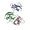
Mass: 65.409 Da / Num. of mol.: 9 / Source method: obtained synthetically / Formula: Zn / Feature type: SUBJECT OF INVESTIGATION
#3: Water ChemComp-HOH / water / Water


Mass: 18.015 Da / Num. of mol.: 55 / Source method: isolated from a natural source / Formula: H2O
Has ligand of interestY

-
Experimental details

-
Experiment

ExperimentMethod: X-RAY DIFFRACTION / Number of used crystals: 1

-
Sample preparation

CrystalDensity Matthews: 2.92 Å3/Da / Density % sol: 57.89 %
Crystal growTemperature: 291.15 K / Method: vapor diffusion, hanging drop / pH: 7
Details: 0.1 M DL-malic acid (pH7.0) and 18% (w/v) polyethylene glycol 3350 (PEG3350)

-
Data collection

DiffractionMean temperature: 100 K / Serial crystal experiment: N
Diffraction sourceSource: SYNCHROTRON / Site: PAL/PLS / Beamline: 11C / Wavelength: 1.283 Å
DetectorType: DECTRIS PILATUS3 6M / Detector: PIXEL / Date: Jun 15, 2022
RadiationProtocol: SINGLE WAVELENGTH / Monochromatic (M) / Laue (L): M / Scattering type: x-ray
Radiation wavelengthWavelength: 1.283 Å / Relative weight: 1
ReflectionResolution: 2.18→29.76 Å / Num. obs: 15341 / % possible obs: 98.04 % / Redundancy: 12.7 % / Biso Wilson estimate: 40.55 Å2 / CC1/2: 0.999 / Rmerge(I) obs: 0.15 / Net I/σ(I): 13.99
Reflection shellResolution: 2.18→2.258 Å / Num. unique obs: 1490 / CC1/2: 0.926

-
Processing

Software
NameVersionClassification
PHENIX1.17.1_3660refinement
XDSdata reduction
XDSdata scaling
PHENIXphasing
RefinementMethod to determine structure: MOLECULAR REPLACEMENT
Starting model: AlphaFold

Resolution: 2.18→29.76 Å / SU ML: 0.334 / Cross valid method: FREE R-VALUE / σ(F): 1.36 / Phase error: 34.6495
Stereochemistry target values: GeoStd + Monomer Library + CDL v1.2
RfactorNum. reflection% reflection
Rfree0.2688 762 4.99 %
Rwork0.2192 14510 -
obs0.2217 15272 98.08 %
Solvent computationShrinkage radii: 0.9 Å / VDW probe radii: 1.11 Å / Solvent model: FLAT BULK SOLVENT MODEL
Displacement parametersBiso mean: 49.78 Å2
Refinement stepCycle: LAST / Resolution: 2.18→29.76 Å
ProteinNucleic acidLigandSolventTotal
Num. atoms1690 0 9 55 1754
Refine LS restraints
Refine-IDTypeDev idealNumber
X-RAY DIFFRACTIONf_bond_d0.00761732
X-RAY DIFFRACTIONf_angle_d0.84442325
X-RAY DIFFRACTIONf_chiral_restr0.0549248
X-RAY DIFFRACTIONf_plane_restr0.0039288
X-RAY DIFFRACTIONf_dihedral_angle_d6.7773225
LS refinement shell
Resolution (Å)Rfactor RfreeNum. reflection RfreeRfactor RworkNum. reflection RworkRefine-ID% reflection obs (%)
2.18-2.350.38921460.36182821X-RAY DIFFRACTION98.05
2.35-2.580.30341500.27582862X-RAY DIFFRACTION97.95
2.58-2.960.37911520.25972876X-RAY DIFFRACTION98.15
2.96-3.720.26851540.22762911X-RAY DIFFRACTION98.17
3.73-29.760.21551600.17193040X-RAY DIFFRACTION98.1

+
About Yorodumi

-
News

-
Feb 9, 2022. New format data for meta-information of EMDB entries

New format data for meta-information of EMDB entries

  • Version 3 of the EMDB header file is now the official format.
  • The previous official version 1.9 will be removed from the archive.

Related info.:EMDB header

External links:wwPDB to switch to version 3 of the EMDB data model

-
Aug 12, 2020. Covid-19 info

Covid-19 info

URL: https://pdbj.org/emnavi/covid19.php

New page: Covid-19 featured information page in EM Navigator.

Related info.:Covid-19 info / Mar 5, 2020. Novel coronavirus structure data

+
Mar 5, 2020. Novel coronavirus structure data

Novel coronavirus structure data

Related info.:Yorodumi Speices / Aug 12, 2020. Covid-19 info

External links:COVID-19 featured content - PDBj / Molecule of the Month (242):Coronavirus Proteases

+
Jan 31, 2019. EMDB accession codes are about to change! (news from PDBe EMDB page)

EMDB accession codes are about to change! (news from PDBe EMDB page)

  • The allocation of 4 digits for EMDB accession codes will soon come to an end. Whilst these codes will remain in use, new EMDB accession codes will include an additional digit and will expand incrementally as the available range of codes is exhausted. The current 4-digit format prefixed with “EMD-” (i.e. EMD-XXXX) will advance to a 5-digit format (i.e. EMD-XXXXX), and so on. It is currently estimated that the 4-digit codes will be depleted around Spring 2019, at which point the 5-digit format will come into force.
  • The EM Navigator/Yorodumi systems omit the EMD- prefix.

Related info.:Q: What is EMD? / ID/Accession-code notation in Yorodumi/EM Navigator

External links:EMDB Accession Codes are Changing Soon! / Contact to PDBj

+
Jul 12, 2017. Major update of PDB

Major update of PDB

  • wwPDB released updated PDB data conforming to the new PDBx/mmCIF dictionary.
  • This is a major update changing the version number from 4 to 5, and with Remediation, in which all the entries are updated.
  • In this update, many items about electron microscopy experimental information are reorganized (e.g. em_software).
  • Now, EM Navigator and Yorodumi are based on the updated data.

External links:wwPDB Remediation / Enriched Model Files Conforming to OneDep Data Standards Now Available in the PDB FTP Archive

-
Yorodumi

Thousand views of thousand structures

  • Yorodumi is a browser for structure data from EMDB, PDB, SASBDB, etc.
  • This page is also the successor to EM Navigator detail page, and also detail information page/front-end page for Omokage search.
  • The word "yorodu" (or yorozu) is an old Japanese word meaning "ten thousand". "mi" (miru) is to see.

Related info.:EMDB / PDB / SASBDB / Comparison of 3 databanks / Yorodumi Search / Aug 31, 2016. New EM Navigator & Yorodumi / Yorodumi Papers / Jmol/JSmol / Function and homology information / Changes in new EM Navigator and Yorodumi

Read more