[English] 日本語
Yorodumi
- PDB-8j9f: Structure of STG-hydrolyzing beta-glucosidase 1 (PSTG1) -

+
Open data


ID or keywords:

Loading...

-
Basic information

Entry
Database: PDB / ID: 8j9f
TitleStructure of STG-hydrolyzing beta-glucosidase 1 (PSTG1)
ComponentsBeta-glucosidase
KeywordsHYDROLASE / GH3 family / sesaminol biosynthesis
Function / homology
Function and homology information


scopolin beta-glucosidase activity / beta-glucosidase activity / beta-glucosidase / carbohydrate metabolic process
Similarity search - Function
Fibronectin type III-like domain / Fibronectin type III-like domain / Fibronectin type III-like domain / Glycoside hydrolase, family 3, active site / Glycosyl hydrolases family 3 active site. / Glycoside hydrolase family 3 C-terminal domain / Glycosyl hydrolase family 3 C-terminal domain / Glycoside hydrolase family 3 C-terminal domain superfamily / Glycoside hydrolase, family 3, N-terminal / Glycoside hydrolase, family 3, N-terminal domain superfamily ...Fibronectin type III-like domain / Fibronectin type III-like domain / Fibronectin type III-like domain / Glycoside hydrolase, family 3, active site / Glycosyl hydrolases family 3 active site. / Glycoside hydrolase family 3 C-terminal domain / Glycosyl hydrolase family 3 C-terminal domain / Glycoside hydrolase family 3 C-terminal domain superfamily / Glycoside hydrolase, family 3, N-terminal / Glycoside hydrolase, family 3, N-terminal domain superfamily / Glycosyl hydrolase family 3 N terminal domain / Glycoside hydrolase superfamily / Immunoglobulin-like fold
Similarity search - Domain/homology
Biological speciesPaenibacillus relictisesami (bacteria)
MethodX-RAY DIFFRACTION / SYNCHROTRON / MOLECULAR REPLACEMENT / Resolution: 2.85 Å
AuthorsYanai, T. / Imaizumi, R. / Takahashi, Y. / Katsumura, E. / Yamamoto, M. / Nakayama, T. / Yamashita, S. / Takeshita, K. / Sakai, N. / Matsuura, H.
Funding support Japan, 2items
OrganizationGrant numberCountry
Japan Society for the Promotion of Science (JSPS)23H05470 Japan
Japan Agency for Medical Research and Development (AMED) Japan
CitationJournal: To be published
Title: Structural insights into a bacterial beta-glucosidase that is capable of degrading sesaminol triglucoside to produce sesaminol
Authors: Yanai, T. / Imaizumi, R. / Nakayama, T. / Yamashita, S.
History
DepositionMay 3, 2023Deposition site: PDBJ / Processing site: PDBJ
Revision 1.0Apr 10, 2024Provider: repository / Type: Initial release

-
Structure visualization

Structure viewerMolecule:
MolmilJmol/JSmol

Downloads & links

-
Assembly

Deposited unit
A: Beta-glucosidase
B: Beta-glucosidase
C: Beta-glucosidase
D: Beta-glucosidase
hetero molecules


Theoretical massNumber of molelcules
Total (without water)346,7987
Polymers346,5224
Non-polymers2763
Water8,845491
1


  • Idetical with deposited unit
  • defined by author
  • Evidence: gel filtration
TypeNameSymmetry operationNumber
identity operation1_555x,y,z1
Buried area13560 Å2
ΔGint-68 kcal/mol
Surface area105810 Å2
2
A: Beta-glucosidase
B: Beta-glucosidase
hetero molecules


Theoretical massNumber of molelcules
Total (without water)173,4454
Polymers173,2612
Non-polymers1842
Water362
TypeNameSymmetry operationNumber
identity operation1_555x,y,z1
Buried area6540 Å2
ΔGint-33 kcal/mol
Surface area53240 Å2
MethodPISA
3
C: Beta-glucosidase
D: Beta-glucosidase
hetero molecules


Theoretical massNumber of molelcules
Total (without water)173,3533
Polymers173,2612
Non-polymers921
Water362
TypeNameSymmetry operationNumber
identity operation1_555x,y,z1
Buried area6240 Å2
ΔGint-34 kcal/mol
Surface area53350 Å2
MethodPISA
Unit cell
Length a, b, c (Å)65.300, 113.250, 158.660
Angle α, β, γ (deg.)71.468, 82.684, 79.830
Int Tables number1
Space group name H-MP1
Noncrystallographic symmetry (NCS)NCS domain:
IDEns-IDDetails
11A
21A
32A
42A
53A
63A
74A
84A
95A
105A
116A
126A

NCS domain segments:

Beg auth comp-ID: ARG / Beg label comp-ID: ARG / End auth comp-ID: GLY / End label comp-ID: GLY / Auth asym-ID: A / Label asym-ID: A / Auth seq-ID: 5 - 753 / Label seq-ID: 32 - 780

Dom-IDComponent-IDEns-ID
111
211
322
422
533
633
744
844
955
1055
1166
1266

NCS ensembles :
IDDetails
1Local NCS retraints between domains: 1 2
2Local NCS retraints between domains: 3 4
3Local NCS retraints between domains: 5 6
4Local NCS retraints between domains: 7 8
5Local NCS retraints between domains: 9 10
6Local NCS retraints between domains: 11 12

-
Components

#1: Protein
Beta-glucosidase /


Mass: 86630.438 Da / Num. of mol.: 4
Source method: isolated from a genetically manipulated source
Source: (gene. exp.) Paenibacillus relictisesami (bacteria) / Gene: PSTG / Production host: Escherichia coli BL21(DE3) (bacteria) / References: UniProt: D6RVX0
#2: Chemical ChemComp-GOL / GLYCEROL / GLYCERIN / PROPANE-1,2,3-TRIOL / Glycerol


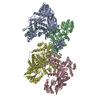
Mass: 92.094 Da / Num. of mol.: 3 / Source method: obtained synthetically / Formula: C3H8O3
#3: Water ChemComp-HOH / water / Water


Mass: 18.015 Da / Num. of mol.: 491 / Source method: isolated from a natural source / Formula: H2O
Has ligand of interestN

-
Experimental details

-
Experiment

ExperimentMethod: X-RAY DIFFRACTION / Number of used crystals: 1

-
Sample preparation

CrystalDensity Matthews: 3.3 Å3/Da / Density % sol: 62.72 %
Crystal growTemperature: 293 K / Method: vapor diffusion, hanging drop / pH: 8 / Details: PEG 3000, imidazole, lithium sulfate

-
Data collection

DiffractionMean temperature: 80 K / Serial crystal experiment: N
Diffraction sourceSource: SYNCHROTRON / Site: SPring-8 / Beamline: BL32XU / Wavelength: 1 Å
DetectorType: DECTRIS EIGER X 9M / Detector: PIXEL / Date: Jun 22, 2021
RadiationProtocol: SINGLE WAVELENGTH / Monochromatic (M) / Laue (L): M / Scattering type: x-ray
Radiation wavelengthWavelength: 1 Å / Relative weight: 1
ReflectionResolution: 2.85→50 Å / Num. obs: 832650 / % possible obs: 99.6 % / Redundancy: 8.46 % / CC1/2: 0.879 / Rrim(I) all: 0.619 / Net I/σ(I): 3.65
Reflection shellResolution: 2.85→3.02 Å / Redundancy: 8.25 % / Mean I/σ(I) obs: 1.33 / Num. unique obs: 131530 / CC1/2: 0.515 / Rrim(I) all: 1.661 / % possible all: 99.8

-
Processing

Software
NameVersionClassification
REFMAC5.8.0405refinement
PHENIX1.20.1-4487refinement
XDSdata reduction
XDSdata scaling
MOLREP11.9.02phasing
RefinementMethod to determine structure: MOLECULAR REPLACEMENT / Resolution: 2.85→47.6 Å / Cor.coef. Fo:Fc: 0.873 / Cor.coef. Fo:Fc free: 0.807 / Cross valid method: FREE R-VALUE / ESU R Free: 0.411
Details: Hydrogens have been added in their riding positions
RfactorNum. reflection% reflectionSelection details
Rfree0.2817 5067 5.146 %Random selection
Rwork0.2317 93394 --
all0.234 ---
obs-98461 99.681 %-
Solvent computationIon probe radii: 0.8 Å / Shrinkage radii: 0.8 Å / VDW probe radii: 1.2 Å / Solvent model: MASK BULK SOLVENT
Displacement parametersBiso mean: 19.839 Å2
Baniso -1Baniso -2Baniso -3
1--1.229 Å2-0.629 Å2-0.115 Å2
2--1.792 Å21.404 Å2
3----0.033 Å2
Refinement stepCycle: LAST / Resolution: 2.85→47.6 Å
ProteinNucleic acidLigandSolventTotal
Num. atoms23104 0 18 491 23613
Refine LS restraints
Refine-IDTypeDev idealDev ideal targetNumber
X-RAY DIFFRACTIONr_bond_refined_d0.0120.01223563
X-RAY DIFFRACTIONr_bond_other_d0.0020.01622454
X-RAY DIFFRACTIONr_angle_refined_deg1.6481.64331864
X-RAY DIFFRACTIONr_angle_other_deg0.8561.56751785
X-RAY DIFFRACTIONr_dihedral_angle_1_deg6.04352981
X-RAY DIFFRACTIONr_dihedral_angle_2_deg3.6455145
X-RAY DIFFRACTIONr_dihedral_angle_3_deg17.346104095
X-RAY DIFFRACTIONr_dihedral_angle_6_deg17.764101052
X-RAY DIFFRACTIONr_chiral_restr0.0720.23552
X-RAY DIFFRACTIONr_gen_planes_refined0.0060.0227617
X-RAY DIFFRACTIONr_gen_planes_other0.0050.025219
X-RAY DIFFRACTIONr_nbd_refined0.2120.24777
X-RAY DIFFRACTIONr_symmetry_nbd_other0.2330.221226
X-RAY DIFFRACTIONr_nbtor_refined0.1860.211495
X-RAY DIFFRACTIONr_symmetry_nbtor_other0.0840.213388
X-RAY DIFFRACTIONr_xyhbond_nbd_refined0.1860.2603
X-RAY DIFFRACTIONr_symmetry_xyhbond_nbd_other0.0670.215
X-RAY DIFFRACTIONr_symmetry_nbd_refined0.2430.219
X-RAY DIFFRACTIONr_nbd_other0.2440.234
X-RAY DIFFRACTIONr_symmetry_xyhbond_nbd_refined0.1150.22
X-RAY DIFFRACTIONr_mcbond_it1.5921.98211942
X-RAY DIFFRACTIONr_mcbond_other1.5921.98111941
X-RAY DIFFRACTIONr_mcangle_it2.6963.55814917
X-RAY DIFFRACTIONr_mcangle_other2.6963.55814918
X-RAY DIFFRACTIONr_scbond_it1.7022.14611621
X-RAY DIFFRACTIONr_scbond_other1.7022.14611622
X-RAY DIFFRACTIONr_scangle_it2.9553.86516947
X-RAY DIFFRACTIONr_scangle_other2.9553.86416948
X-RAY DIFFRACTIONr_lrange_it5.63923.59196861
X-RAY DIFFRACTIONr_lrange_other5.63823.60596777
X-RAY DIFFRACTIONr_ncsr_local_group_10.120.0522514
X-RAY DIFFRACTIONr_ncsr_local_group_20.1250.0522492
X-RAY DIFFRACTIONr_ncsr_local_group_30.1220.0522529
X-RAY DIFFRACTIONr_ncsr_local_group_40.1230.0522447
X-RAY DIFFRACTIONr_ncsr_local_group_50.1250.0522443
X-RAY DIFFRACTIONr_ncsr_local_group_60.120.0522755
Refine LS restraints NCS
Ens-IDDom-IDAuth asym-IDRefine-IDTypeRms dev position (Å)Weight position
11AX-RAY DIFFRACTIONLocal ncs0.119970.05007
12AX-RAY DIFFRACTIONLocal ncs0.119970.05007
23AX-RAY DIFFRACTIONLocal ncs0.125280.05007
24AX-RAY DIFFRACTIONLocal ncs0.125280.05007
35AX-RAY DIFFRACTIONLocal ncs0.122190.05007
36AX-RAY DIFFRACTIONLocal ncs0.122190.05007
47AX-RAY DIFFRACTIONLocal ncs0.123190.05007
48AX-RAY DIFFRACTIONLocal ncs0.123190.05007
59AX-RAY DIFFRACTIONLocal ncs0.124850.05007
510AX-RAY DIFFRACTIONLocal ncs0.124850.05007
611AX-RAY DIFFRACTIONLocal ncs0.119950.05007
612AX-RAY DIFFRACTIONLocal ncs0.119950.05007
LS refinement shell
Resolution (Å)Rfactor RfreeNum. reflection RfreeRfactor RworkNum. reflection RworkRefine-ID% reflection obs (%)
2.85-2.9240.3463770.3116904X-RAY DIFFRACTION99.8492
2.924-3.0030.3093350.296730X-RAY DIFFRACTION99.774
3.003-3.090.3583570.2896546X-RAY DIFFRACTION99.7976
3.09-3.1850.3363470.2746312X-RAY DIFFRACTION99.7155
3.185-3.2890.3313130.2666147X-RAY DIFFRACTION99.4458
3.289-3.4040.3143260.2635930X-RAY DIFFRACTION99.1442
3.404-3.5310.3073060.2465734X-RAY DIFFRACTION99.5222
3.531-3.6750.273320.2315522X-RAY DIFFRACTION99.8295
3.675-3.8370.2672660.2265323X-RAY DIFFRACTION99.7679
3.837-4.0230.2843090.2165019X-RAY DIFFRACTION99.794
4.023-4.2390.2472780.194796X-RAY DIFFRACTION99.8229
4.239-4.4940.2252490.1764565X-RAY DIFFRACTION99.8548
4.494-4.8010.232360.1784318X-RAY DIFFRACTION99.8028
4.801-5.1820.2261810.1774023X-RAY DIFFRACTION99.8812
5.182-5.6690.2612050.2033690X-RAY DIFFRACTION99.7184
5.669-6.3270.2661650.2313344X-RAY DIFFRACTION99.7725
6.327-7.2850.2991680.242945X-RAY DIFFRACTION99.8717
7.285-8.8710.2191380.2012479X-RAY DIFFRACTION99.7713
8.871-12.3330.2331150.1991947X-RAY DIFFRACTION99.8064
12.333-47.60.317640.2621120X-RAY DIFFRACTION98.5845

+
About Yorodumi

-
News

-
Feb 9, 2022. New format data for meta-information of EMDB entries

New format data for meta-information of EMDB entries

  • Version 3 of the EMDB header file is now the official format.
  • The previous official version 1.9 will be removed from the archive.

Related info.:EMDB header

External links:wwPDB to switch to version 3 of the EMDB data model

-
Aug 12, 2020. Covid-19 info

Covid-19 info

URL: https://pdbj.org/emnavi/covid19.php

New page: Covid-19 featured information page in EM Navigator.

Related info.:Covid-19 info / Mar 5, 2020. Novel coronavirus structure data

+
Mar 5, 2020. Novel coronavirus structure data

Novel coronavirus structure data

Related info.:Yorodumi Speices / Aug 12, 2020. Covid-19 info

External links:COVID-19 featured content - PDBj / Molecule of the Month (242):Coronavirus Proteases

+
Jan 31, 2019. EMDB accession codes are about to change! (news from PDBe EMDB page)

EMDB accession codes are about to change! (news from PDBe EMDB page)

  • The allocation of 4 digits for EMDB accession codes will soon come to an end. Whilst these codes will remain in use, new EMDB accession codes will include an additional digit and will expand incrementally as the available range of codes is exhausted. The current 4-digit format prefixed with “EMD-” (i.e. EMD-XXXX) will advance to a 5-digit format (i.e. EMD-XXXXX), and so on. It is currently estimated that the 4-digit codes will be depleted around Spring 2019, at which point the 5-digit format will come into force.
  • The EM Navigator/Yorodumi systems omit the EMD- prefix.

Related info.:Q: What is EMD? / ID/Accession-code notation in Yorodumi/EM Navigator

External links:EMDB Accession Codes are Changing Soon! / Contact to PDBj

+
Jul 12, 2017. Major update of PDB

Major update of PDB

  • wwPDB released updated PDB data conforming to the new PDBx/mmCIF dictionary.
  • This is a major update changing the version number from 4 to 5, and with Remediation, in which all the entries are updated.
  • In this update, many items about electron microscopy experimental information are reorganized (e.g. em_software).
  • Now, EM Navigator and Yorodumi are based on the updated data.

External links:wwPDB Remediation / Enriched Model Files Conforming to OneDep Data Standards Now Available in the PDB FTP Archive

-
Yorodumi

Thousand views of thousand structures

  • Yorodumi is a browser for structure data from EMDB, PDB, SASBDB, etc.
  • This page is also the successor to EM Navigator detail page, and also detail information page/front-end page for Omokage search.
  • The word "yorodu" (or yorozu) is an old Japanese word meaning "ten thousand". "mi" (miru) is to see.

Related info.:EMDB / PDB / SASBDB / Comparison of 3 databanks / Yorodumi Search / Aug 31, 2016. New EM Navigator & Yorodumi / Yorodumi Papers / Jmol/JSmol / Function and homology information / Changes in new EM Navigator and Yorodumi

Read more