[English] 日本語
Yorodumi
- PDB-8j79: Bovine Xanthine Oxidoreductase Crystallized with oxypurinol -

+
Open data


ID or keywords:

Loading...

-
Basic information

Entry
Database: PDB / ID: 8j79
TitleBovine Xanthine Oxidoreductase Crystallized with oxypurinol
ComponentsXanthine dehydrogenase/oxidase
KeywordsOXIDOREDUCTASE / xanthine oxidoreductase / xanthine oxidase / xanthine dehydrogenase
Function / homology
Function and homology information


xanthine dehydrogenase complex / xanthine dehydrogenase / xanthine oxidase / xanthine oxidase activity / xanthine catabolic process / xanthine dehydrogenase activity / molybdenum ion binding / molybdopterin cofactor binding / FAD binding / 2 iron, 2 sulfur cluster binding ...xanthine dehydrogenase complex / xanthine dehydrogenase / xanthine oxidase / xanthine oxidase activity / xanthine catabolic process / xanthine dehydrogenase activity / molybdenum ion binding / molybdopterin cofactor binding / FAD binding / 2 iron, 2 sulfur cluster binding / peroxisome / flavin adenine dinucleotide binding / iron ion binding / protein homodimerization activity / extracellular space
Similarity search - Function
Xanthine dehydrogenase, small subunit / Oxidoreductase, molybdopterin binding site / Eukaryotic molybdopterin oxidoreductases signature. / CO dehydrogenase flavoprotein C-terminal domain / Molybdopterin dehydrogenase, FAD-binding / CO dehydrogenase flavoprotein, C-terminal / CO dehydrogenase flavoprotein, C-terminal domain superfamily / FAD binding domain in molybdopterin dehydrogenase / CO dehydrogenase flavoprotein C-terminal domain / Aldehyde oxidase/xanthine dehydrogenase ...Xanthine dehydrogenase, small subunit / Oxidoreductase, molybdopterin binding site / Eukaryotic molybdopterin oxidoreductases signature. / CO dehydrogenase flavoprotein C-terminal domain / Molybdopterin dehydrogenase, FAD-binding / CO dehydrogenase flavoprotein, C-terminal / CO dehydrogenase flavoprotein, C-terminal domain superfamily / FAD binding domain in molybdopterin dehydrogenase / CO dehydrogenase flavoprotein C-terminal domain / Aldehyde oxidase/xanthine dehydrogenase / Aldehyde oxidase/xanthine dehydrogenase, second molybdopterin binding domain / Molybdopterin cofactor-binding domain / Aldehyde oxidase and xanthine dehydrogenase, a/b hammerhead domain / Aldehyde oxidase/xanthine dehydrogenase, a/b hammerhead / [2Fe-2S]-binding / Aldehyde oxidase/xanthine dehydrogenase, first molybdopterin binding domain / Aldehyde oxidase/xanthine dehydrogenase, a/b hammerhead superfamily / [2Fe-2S]-binding domain superfamily / Aldehyde oxidase/xanthine dehydrogenase, molybdopterin binding domain superfamily / [2Fe-2S] binding domain / Molybdopterin cofactor-binding domain / Aldehyde oxidase and xanthine dehydrogenase, a/b hammerhead domain / FAD-binding, type PCMH, subdomain 1 / 2Fe-2S ferredoxin, iron-sulphur binding site / 2Fe-2S ferredoxin-type iron-sulfur binding region signature. / FAD-binding domain, PCMH-type / PCMH-type FAD-binding domain profile. / FAD-binding, type PCMH, subdomain 2 / 2Fe-2S iron-sulfur cluster binding domain / FAD-binding, type PCMH-like superfamily / Beta-grasp domain superfamily / 2Fe-2S ferredoxin-type iron-sulfur binding domain profile. / 2Fe-2S ferredoxin-type iron-sulfur binding domain / 2Fe-2S ferredoxin-like superfamily
Similarity search - Domain/homology
Oxypurinol / CARBONATE ION / FLAVIN-ADENINE DINUCLEOTIDE / FE2/S2 (INORGANIC) CLUSTER / DIOXOTHIOMOLYBDENUM(VI) ION / Chem-MTE / Xanthine dehydrogenase/oxidase
Similarity search - Component
Biological speciesBos taurus (cattle)
MethodX-RAY DIFFRACTION / SYNCHROTRON / MOLECULAR REPLACEMENT / Resolution: 1.99 Å
AuthorsOkamoto, K. / Sekine, M.
Funding support Japan, 1items
OrganizationGrant numberCountry
Japan Science and Technology15H04702 Japan
CitationJournal: To Be Published
Title: Bovine Xanthine Oxidoreductase Crystallized with oxypurinol
Authors: Okamoto, K. / Sekine, M.
History
DepositionApr 27, 2023Deposition site: PDBJ / Processing site: PDBJ
Revision 1.0May 24, 2023Provider: repository / Type: Initial release
Revision 1.1May 29, 2024Group: Data collection / Category: chem_comp_atom / chem_comp_bond

-
Structure visualization

Structure viewerMolecule:
MolmilJmol/JSmol

Downloads & links

-
Assembly

Deposited unit
A: Xanthine dehydrogenase/oxidase
B: Xanthine dehydrogenase/oxidase
hetero molecules


Theoretical massNumber of molelcules
Total (without water)298,46426
Polymers293,9402
Non-polymers4,52424
Water32,6971815
1


  • Idetical with deposited unit
  • defined by author&software
  • Evidence: gel filtration
TypeNameSymmetry operationNumber
identity operation1_555x,y,z1
Buried area14870 Å2
ΔGint-147 kcal/mol
Surface area85540 Å2
MethodPISA
Unit cell
Length a, b, c (Å)167.678, 123.470, 150.059
Angle α, β, γ (deg.)90.00, 90.89, 90.00
Int Tables number5
Space group name H-MC121

-
Components

-
Protein , 1 types, 2 molecules AB

#1: Protein Xanthine dehydrogenase/oxidase


Mass: 146970.078 Da / Num. of mol.: 2
Source method: isolated from a genetically manipulated source
Source: (gene. exp.) Bos taurus (cattle) / Gene: XDH / Production host: Bos taurus (cattle)
References: UniProt: P80457, xanthine dehydrogenase, xanthine oxidase

-
Non-polymers , 9 types, 1839 molecules

#2: Chemical
ChemComp-FES / FE2/S2 (INORGANIC) CLUSTER


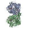
Mass: 175.820 Da / Num. of mol.: 4 / Source method: obtained synthetically / Formula: Fe2S2
#3: Chemical ChemComp-MTE / PHOSPHONIC ACIDMONO-(2-AMINO-5,6-DIMERCAPTO-4-OXO-3,7,8A,9,10,10A-HEXAHYDRO-4H-8-OXA-1,3,9,10-TETRAAZA-ANTHRACEN-7-YLMETHYL)ESTER


Mass: 395.352 Da / Num. of mol.: 2 / Source method: obtained synthetically / Formula: C10H14N5O6PS2
#4: Chemical ChemComp-MOS / DIOXOTHIOMOLYBDENUM(VI) ION


Mass: 161.012 Da / Num. of mol.: 2 / Source method: obtained synthetically / Formula: HMoO2S
#5: Chemical ChemComp-FAD / FLAVIN-ADENINE DINUCLEOTIDE


Mass: 785.550 Da / Num. of mol.: 2 / Source method: obtained synthetically / Formula: C27H33N9O15P2 / Comment: FAD*YM
#6: Chemical ChemComp-CO3 / CARBONATE ION


Mass: 60.009 Da / Num. of mol.: 2 / Source method: obtained synthetically / Formula: CO3
#7: Chemical
ChemComp-CA / CALCIUM ION


Mass: 40.078 Da / Num. of mol.: 4 / Source method: obtained synthetically / Formula: Ca
#8: Chemical ChemComp-141 / Oxypurinol / Alloxanthine


Mass: 152.111 Da / Num. of mol.: 2 / Source method: obtained synthetically / Formula: C5H4N4O2 / Comment: inhibitor*YM
#9: Chemical
ChemComp-GOL / GLYCEROL / GLYCERIN / PROPANE-1,2,3-TRIOL


Mass: 92.094 Da / Num. of mol.: 6 / Source method: obtained synthetically / Formula: C3H8O3
#10: Water ChemComp-HOH / water


Mass: 18.015 Da / Num. of mol.: 1815 / Source method: isolated from a natural source / Formula: H2O

-
Details

Has ligand of interestN

-
Experimental details

-
Experiment

ExperimentMethod: X-RAY DIFFRACTION / Number of used crystals: 1

-
Sample preparation

CrystalDensity Matthews: 2.73 Å3/Da / Density % sol: 54.92 %
Crystal growTemperature: 293 K / Method: batch mode
Details: The enzyme solution was exchanged buffer into 50 mM Tris-HCl pH 7.4 and diluted with 50 mM Tris-HCl pH 7.4, 5 mM DTT, 100 micro M oxypurinol, and 30% glycerol to a concentration of 8.6 mg/ml. ...Details: The enzyme solution was exchanged buffer into 50 mM Tris-HCl pH 7.4 and diluted with 50 mM Tris-HCl pH 7.4, 5 mM DTT, 100 micro M oxypurinol, and 30% glycerol to a concentration of 8.6 mg/ml. The precipitant solution contained 50 mM potassium phosphate pH 6.0, 5 mM DTT, 0.2 mM EDTA, 100 micro M oxypurinol, 30% glycerol and 14%-15% PEG 4000. 10 micro L enzyme solution and 10 micro L reservoir solution were mixed on siliconized glass plates and kept in the dark at 20 C for 5 days.

-
Data collection

DiffractionMean temperature: 100 K / Serial crystal experiment: N
Diffraction sourceSource: SYNCHROTRON / Site: SPring-8 / Beamline: BL44XU / Wavelength: 0.9 Å
DetectorType: DECTRIS EIGER X 16M / Detector: PIXEL / Date: Jul 21, 2020
RadiationProtocol: SINGLE WAVELENGTH / Monochromatic (M) / Laue (L): M / Scattering type: x-ray
Radiation wavelengthWavelength: 0.9 Å / Relative weight: 1
ReflectionResolution: 1.99→48.43 Å / Num. obs: 210046 / % possible obs: 99.7 % / Redundancy: 5 % / CC1/2: 0.996 / Net I/σ(I): 10.6
Reflection shellResolution: 1.99→2.11 Å / Num. unique obs: 33646 / CC1/2: 0.843

-
Processing

Software
NameVersionClassification
REFMAC5.8.0258refinement
XDSdata reduction
XDSdata scaling
MOLREPphasing
RefinementMethod to determine structure: MOLECULAR REPLACEMENT / Resolution: 1.99→47.72 Å / Cor.coef. Fo:Fc: 0.962 / Cor.coef. Fo:Fc free: 0.949 / SU B: 3.669 / SU ML: 0.1 / Cross valid method: THROUGHOUT / ESU R: 0.147 / ESU R Free: 0.132 / Stereochemistry target values: MAXIMUM LIKELIHOOD / Details: HYDROGENS HAVE BEEN ADDED IN THE RIDING POSITIONS
RfactorNum. reflection% reflectionSelection details
Rfree0.19667 10629 5.1 %RANDOM
Rwork0.16346 ---
obs0.16515 199435 99.74 %-
Solvent computationIon probe radii: 0.8 Å / Shrinkage radii: 0.8 Å / VDW probe radii: 1.2 Å / Solvent model: MASK
Displacement parametersBiso mean: 29.898 Å2
Baniso -1Baniso -2Baniso -3
1-0 Å20 Å20 Å2
2--0 Å20 Å2
3----0 Å2
Refinement stepCycle: 1 / Resolution: 1.99→47.72 Å
ProteinNucleic acidLigandSolventTotal
Num. atoms19980 0 248 1815 22043
Refine LS restraints
Refine-IDTypeDev idealDev ideal targetNumber
X-RAY DIFFRACTIONr_bond_refined_d0.010.01320690
X-RAY DIFFRACTIONr_bond_other_d0.0010.01719231
X-RAY DIFFRACTIONr_angle_refined_deg1.6231.64928022
X-RAY DIFFRACTIONr_angle_other_deg1.3811.57244710
X-RAY DIFFRACTIONr_dihedral_angle_1_deg6.97552565
X-RAY DIFFRACTIONr_dihedral_angle_2_deg33.82322.177992
X-RAY DIFFRACTIONr_dihedral_angle_3_deg13.276153521
X-RAY DIFFRACTIONr_dihedral_angle_4_deg15.52915122
X-RAY DIFFRACTIONr_chiral_restr0.0790.22720
X-RAY DIFFRACTIONr_gen_planes_refined0.0080.0222971
X-RAY DIFFRACTIONr_gen_planes_other0.0010.024271
X-RAY DIFFRACTIONr_nbd_refined
X-RAY DIFFRACTIONr_nbd_other
X-RAY DIFFRACTIONr_nbtor_refined
X-RAY DIFFRACTIONr_nbtor_other
X-RAY DIFFRACTIONr_xyhbond_nbd_refined
X-RAY DIFFRACTIONr_xyhbond_nbd_other
X-RAY DIFFRACTIONr_metal_ion_refined
X-RAY DIFFRACTIONr_metal_ion_other
X-RAY DIFFRACTIONr_symmetry_vdw_refined
X-RAY DIFFRACTIONr_symmetry_vdw_other
X-RAY DIFFRACTIONr_symmetry_hbond_refined
X-RAY DIFFRACTIONr_symmetry_hbond_other
X-RAY DIFFRACTIONr_symmetry_metal_ion_refined
X-RAY DIFFRACTIONr_symmetry_metal_ion_other
X-RAY DIFFRACTIONr_mcbond_it2.3122.95410284
X-RAY DIFFRACTIONr_mcbond_other2.3122.95410283
X-RAY DIFFRACTIONr_mcangle_it3.0654.42112841
X-RAY DIFFRACTIONr_mcangle_other3.0654.42112842
X-RAY DIFFRACTIONr_scbond_it3.0953.31410406
X-RAY DIFFRACTIONr_scbond_other3.0953.31410407
X-RAY DIFFRACTIONr_scangle_it
X-RAY DIFFRACTIONr_scangle_other4.544.8315174
X-RAY DIFFRACTIONr_long_range_B_refined5.80636.05923680
X-RAY DIFFRACTIONr_long_range_B_other5.69735.59123172
X-RAY DIFFRACTIONr_rigid_bond_restr
X-RAY DIFFRACTIONr_sphericity_free
X-RAY DIFFRACTIONr_sphericity_bonded
LS refinement shellResolution: 1.99→2.036 Å
RfactorNum. reflection% reflection
Rfree0.279 805 -
Rwork0.241 14500 -
obs--98.26 %

+
About Yorodumi

-
News

-
Feb 9, 2022. New format data for meta-information of EMDB entries

New format data for meta-information of EMDB entries

  • Version 3 of the EMDB header file is now the official format.
  • The previous official version 1.9 will be removed from the archive.

Related info.:EMDB header

External links:wwPDB to switch to version 3 of the EMDB data model

-
Aug 12, 2020. Covid-19 info

Covid-19 info

URL: https://pdbjlvh1.pdbj.org/emnavi/covid19.php

New page: Covid-19 featured information page in EM Navigator.

Related info.:Covid-19 info / Mar 5, 2020. Novel coronavirus structure data

+
Mar 5, 2020. Novel coronavirus structure data

Novel coronavirus structure data

Related info.:Yorodumi Speices / Aug 12, 2020. Covid-19 info

External links:COVID-19 featured content - PDBj / Molecule of the Month (242):Coronavirus Proteases

+
Jan 31, 2019. EMDB accession codes are about to change! (news from PDBe EMDB page)

EMDB accession codes are about to change! (news from PDBe EMDB page)

  • The allocation of 4 digits for EMDB accession codes will soon come to an end. Whilst these codes will remain in use, new EMDB accession codes will include an additional digit and will expand incrementally as the available range of codes is exhausted. The current 4-digit format prefixed with “EMD-” (i.e. EMD-XXXX) will advance to a 5-digit format (i.e. EMD-XXXXX), and so on. It is currently estimated that the 4-digit codes will be depleted around Spring 2019, at which point the 5-digit format will come into force.
  • The EM Navigator/Yorodumi systems omit the EMD- prefix.

Related info.:Q: What is EMD? / ID/Accession-code notation in Yorodumi/EM Navigator

External links:EMDB Accession Codes are Changing Soon! / Contact to PDBj

+
Jul 12, 2017. Major update of PDB

Major update of PDB

  • wwPDB released updated PDB data conforming to the new PDBx/mmCIF dictionary.
  • This is a major update changing the version number from 4 to 5, and with Remediation, in which all the entries are updated.
  • In this update, many items about electron microscopy experimental information are reorganized (e.g. em_software).
  • Now, EM Navigator and Yorodumi are based on the updated data.

External links:wwPDB Remediation / Enriched Model Files Conforming to OneDep Data Standards Now Available in the PDB FTP Archive

-
Yorodumi

Thousand views of thousand structures

  • Yorodumi is a browser for structure data from EMDB, PDB, SASBDB, etc.
  • This page is also the successor to EM Navigator detail page, and also detail information page/front-end page for Omokage search.
  • The word "yorodu" (or yorozu) is an old Japanese word meaning "ten thousand". "mi" (miru) is to see.

Related info.:EMDB / PDB / SASBDB / Comparison of 3 databanks / Yorodumi Search / Aug 31, 2016. New EM Navigator & Yorodumi / Yorodumi Papers / Jmol/JSmol / Function and homology information / Changes in new EM Navigator and Yorodumi

Read more