 Movie
Movie Controller

[English] 日本語
 Yorodumi
- PDB-8j6h: Structure and allosteric regulation of the inosine 5'-monophospha... -
+ Open data

- Basic information
| Entry | Database: PDB / ID: 8j6h | |||||||||
|---|---|---|---|---|---|---|---|---|---|---|
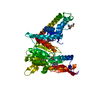
| Title | Structure and allosteric regulation of the inosine 5'-monophosphate-specific phosphatase ISN1 from Saccharomyces cerevisiae | |||||||||
 Components Components | IMP-specific 5'-nucleotidase 1 | |||||||||
 Keywords Keywords | HYDROLASE / hydrolysis / phosphatase | |||||||||
| Function / homology |  Function and homology information Function and homology informationnicotinamide riboside biosynthetic process / nicotinic acid riboside biosynthetic process / inosine salvage / IMP-specific 5'-nucleotidase / 5'-nucleotidase activity / nucleotide metabolic process / magnesium ion binding / ATP binding Similarity search - Function | |||||||||
| Biological species |  | |||||||||
| Method |  X-RAY DIFFRACTION / SYNCHROTRON / SAD / Resolution: 2.44074489024 Å X-RAY DIFFRACTION / SYNCHROTRON / SAD / Resolution: 2.44074489024 Å | |||||||||
 Authors Authors | Byun, S.J. / Rhee, S. | |||||||||
| Funding support |  Korea, Republic Of, 2items Korea, Republic Of, 2items
| |||||||||
 Citation Citation | Journal: Febs J. / Year: 2024 Title: Structure, cooperativity and inhibition of the inosine 5'-monophosphate-specific phosphatase from Saccharomyces cerevisiae. Authors: Byun, S. / Park, C. / Suh, J.Y. / Witte, C.P. / Rhee, S. | |||||||||
| History |
|
- Structure visualization
| Structure viewer | Molecule: MolmilJmol/JSmol |
|---|
- Downloads & links
-Download
| PDBx/mmCIF format | 8j6h.cif.gz | 411.1 KB | Display | PDBx/mmCIF format |
|---|---|---|---|---|
| PDB format | pdb8j6h.ent.gz | 269.6 KB | Display | PDB format |
| PDBx/mmJSON format | 8j6h.json.gz | Tree view | PDBx/mmJSON format | |
| Others |  Other downloads Other downloads |
-Validation report
| Arichive directory | https://data.pdbj.org/pub/pdb/validation_reports/j6/8j6hftp://data.pdbj.org/pub/pdb/validation_reports/j6/8j6h | HTTPS FTP |
|---|
-Related structure data
-Links
 PDBj
PDBj- Assembly
| Deposited unit | 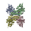
| ||||||||||||
|---|---|---|---|---|---|---|---|---|---|---|---|---|---|
| 1 |
| ||||||||||||
| Unit cell |
|
-Components
| #1: Protein | Mass: 52379.172 Da / Num. of mol.: 4 Source method: isolated from a genetically manipulated source Source: (gene. exp.) Gene: ISN1, YOR155C, O3548 / Variant: DELTA4R / Plasmid: pET41a / Production host:  #2: Chemical | ChemComp-NOS / 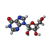  Mass: 268.226 Da / Num. of mol.: 4 / Source method: obtained synthetically / Formula: C10H12N4O5 / Feature type: SUBJECT OF INVESTIGATION Mass: 268.226 Da / Num. of mol.: 4 / Source method: obtained synthetically / Formula: C10H12N4O5 / Feature type: SUBJECT OF INVESTIGATION#3: Water | ChemComp-HOH / |  Mass: 18.015 Da / Num. of mol.: 44 / Source method: isolated from a natural source / Formula: H2O Mass: 18.015 Da / Num. of mol.: 44 / Source method: isolated from a natural source / Formula: H2OHas ligand of interest | Y | Has protein modification | Y | |
|---|
-Experimental details
-Experiment
| Experiment | Method: X-RAY DIFFRACTION / Number of used crystals: 1 |
|---|
- Sample preparation
| Crystal | Density Matthews: 2.41 Å3/Da / Density % sol: 48.93 % |
|---|---|
| Crystal grow | Temperature: 295.15 K / Method: vapor diffusion, sitting drop / pH: 4.6 / Details: 0.26M ammonium dihydrogen phosphate, 35% glycerol |
-Data collection
| Diffraction | Mean temperature: 100 K / Serial crystal experiment: N |
|---|---|
| Diffraction source | Source: SYNCHROTRON / Site: PAL/PLS / Beamline: 11C / Wavelength: 0.97926 Å |
| Detector | Type: DECTRIS PILATUS3 6M / Detector: PIXEL / Date: Jul 30, 2020 |
| Radiation | Monochromator: M / Protocol: SINGLE WAVELENGTH / Monochromatic (M) / Laue (L): M / Scattering type: x-ray |
| Radiation wavelength | Wavelength: 0.97926 Å / Relative weight: 1 |
| Reflection | Resolution: 2.44→50 Å / Num. obs: 74898 / % possible obs: 99.5 % / Redundancy: 6.4 % / Biso Wilson estimate: 55.7092581885 Å2 / CC1/2: 0.99 / Rmerge(I) obs: 0.11 / Net I/σ(I): 9.34 |
| Reflection shell | Resolution: 2.45→2.54 Å / Redundancy: 5.5 % / Num. unique obs: 7175 / CC1/2: 0.48 / % possible all: 96.5 |
- Processing
| Software |
| |||||||||||||||||||||||||||||||||||||||||||||||||||||||||||||||||||||||||||||||||||||||||||||||||||||||||
|---|---|---|---|---|---|---|---|---|---|---|---|---|---|---|---|---|---|---|---|---|---|---|---|---|---|---|---|---|---|---|---|---|---|---|---|---|---|---|---|---|---|---|---|---|---|---|---|---|---|---|---|---|---|---|---|---|---|---|---|---|---|---|---|---|---|---|---|---|---|---|---|---|---|---|---|---|---|---|---|---|---|---|---|---|---|---|---|---|---|---|---|---|---|---|---|---|---|---|---|---|---|---|---|---|---|---|
| Refinement | Method to determine structure: SAD / Resolution: 2.44074489024→47.0886773127 Å / SU ML: 0.40257673769 / Cross valid method: FREE R-VALUE / σ(F): 0.239701093081 / Phase error: 32.3227285541
| |||||||||||||||||||||||||||||||||||||||||||||||||||||||||||||||||||||||||||||||||||||||||||||||||||||||||
| Solvent computation | Shrinkage radii: 0.9 Å / VDW probe radii: 1.11 Å / Solvent model: FLAT BULK SOLVENT MODEL | |||||||||||||||||||||||||||||||||||||||||||||||||||||||||||||||||||||||||||||||||||||||||||||||||||||||||
| Displacement parameters | Biso mean: 66.3725873448 Å2 | |||||||||||||||||||||||||||||||||||||||||||||||||||||||||||||||||||||||||||||||||||||||||||||||||||||||||
| Refinement step | Cycle: LAST / Resolution: 2.44074489024→47.0886773127 Å
| |||||||||||||||||||||||||||||||||||||||||||||||||||||||||||||||||||||||||||||||||||||||||||||||||||||||||
| Refine LS restraints |
| |||||||||||||||||||||||||||||||||||||||||||||||||||||||||||||||||||||||||||||||||||||||||||||||||||||||||
| LS refinement shell |
|
