 Movie
Movie Controller

[English] 日本語
 Yorodumi
- PDB-8j5b: Crystal structure of P domain from norovirus GI.4 capsid protein. -
+ Open data

- Basic information
| Entry | Database: PDB / ID: 8j5b | ||||||
|---|---|---|---|---|---|---|---|
| Title | Crystal structure of P domain from norovirus GI.4 capsid protein. | ||||||
 Components Components | (Capsid protein) x 3 | ||||||
 Keywords Keywords | VIRAL PROTEIN / norovirus / p-domain / capsid | ||||||
| Function / homology | Calicivirus coat protein C-terminal / Calicivirus coat protein C-terminal / Calicivirus coat protein / Calicivirus coat protein / virion component / Picornavirus/Calicivirus coat protein / Viral coat protein subunit / host cell cytoplasm / Capsid protein Function and homology information Function and homology information | ||||||
| Biological species |  Norovirus Hu/GI.4/S50/2008/Lilla Edet/Sweden Norovirus Hu/GI.4/S50/2008/Lilla Edet/Sweden | ||||||
| Method |  X-RAY DIFFRACTION / SYNCHROTRON / MOLECULAR REPLACEMENT / Resolution: 1.89 Å X-RAY DIFFRACTION / SYNCHROTRON / MOLECULAR REPLACEMENT / Resolution: 1.89 Å | ||||||
 Authors Authors | Katsura, K. / Sakai, N. / Hasegawa, K. / Kimura-Someya, T. / Shirouzu, M. | ||||||
| Funding support |  Japan, 1items Japan, 1items
| ||||||
 Citation Citation | Journal: To Be Published Title: Crystal structure of P domain from norovirus GI.4 capsid protein. Authors: Katsura, K. | ||||||
| History |
|
- Structure visualization
| Structure viewer | Molecule: MolmilJmol/JSmol |
|---|
- Downloads & links
-Download
| PDBx/mmCIF format | 8j5b.cif.gz | 243.6 KB | Display | PDBx/mmCIF format |
|---|---|---|---|---|
| PDB format | pdb8j5b.ent.gz | 192.3 KB | Display | PDB format |
| PDBx/mmJSON format | 8j5b.json.gz | Tree view | PDBx/mmJSON format | |
| Others |  Other downloads Other downloads |
-Validation report
| Summary document | 8j5b_validation.pdf.gz | 454.5 KB | Display | wwPDB validaton report |
|---|---|---|---|---|
| Full document | 8j5b_full_validation.pdf.gz | 468.3 KB | Display | |
| Data in XML | 8j5b_validation.xml.gz | 46.1 KB | Display | |
| Data in CIF | 8j5b_validation.cif.gz | 66 KB | Display | |
| Arichive directory | https://data.pdbj.org/pub/pdb/validation_reports/j5/8j5bftp://data.pdbj.org/pub/pdb/validation_reports/j5/8j5b | HTTPS FTP |
-Related structure data
| Similar structure data |
|---|
-Links
 PDBj
PDBj
- Assembly
| Deposited unit | 
| ||||||||
|---|---|---|---|---|---|---|---|---|---|
| 1 | 
| ||||||||
| 2 | 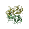
| ||||||||
| Unit cell |
|
-Components
| #1: Protein | Mass: 33895.895 Da / Num. of mol.: 1 Source method: isolated from a genetically manipulated source Source: (gene. exp.) Norovirus Hu/GI.4/S50/2008/Lilla Edet/SwedenGene: ORF2 / Production host:  | ||||
|---|---|---|---|---|---|
| #2: Protein | Mass: 32756.562 Da / Num. of mol.: 2 Source method: isolated from a genetically manipulated source Source: (gene. exp.) Norovirus Hu/GI.4/S50/2008/Lilla Edet/SwedenGene: ORF2 / Production host: #3: Protein | | Mass: 33153.984 Da / Num. of mol.: 1 Source method: isolated from a genetically manipulated source Source: (gene. exp.) Norovirus Hu/GI.4/S50/2008/Lilla Edet/SwedenGene: ORF2 / Production host: #4: Water | ChemComp-HOH / |  Mass: 18.015 Da / Num. of mol.: 441 / Source method: isolated from a natural source / Formula: H2O Mass: 18.015 Da / Num. of mol.: 441 / Source method: isolated from a natural source / Formula: H2O |
-Experimental details
-Experiment
| Experiment | Method: X-RAY DIFFRACTION / Number of used crystals: 1 |
|---|
- Sample preparation
| Crystal | Density Matthews: 2.4 Å3/Da / Density % sol: 48.71 % |
|---|---|
| Crystal grow | Temperature: 293.15 K / Method: vapor diffusion, sitting drop / pH: 5.6 Details: 0.1M tri-Sodium citrate (pH5.6), 20%(v/v) 2-Propanol , 20%(w/v) Polyethylene glycol 4,000 |
-Data collection
| Diffraction | Mean temperature: 93 K / Serial crystal experiment: N |
|---|---|
| Diffraction source | Source: SYNCHROTRON / Site: SPring-8 / Beamline: BL26B2 / Wavelength: 1 Å |
| Detector | Type: MARMOSAIC 225 mm CCD / Detector: CCD / Date: Dec 20, 2016 |
| Radiation | Protocol: SINGLE WAVELENGTH / Monochromatic (M) / Laue (L): M / Scattering type: x-ray |
| Radiation wavelength | Wavelength: 1 Å / Relative weight: 1 |
| Reflection | Resolution: 1.89→49.8 Å / Num. obs: 90970 / % possible obs: 95.9 % / Redundancy: 2.31 % / Biso Wilson estimate: 20.92 Å2 / CC1/2: 0.99 / Rrim(I) all: 0.128 / Rsym value: 0.097 / Net I/σ(I): 6.98 |
| Reflection shell | Resolution: 1.89→2.01 Å / Redundancy: 2.29 % / Mean I/σ(I) obs: 1.54 / Num. unique obs: 14442 / CC1/2: 0.849 / Rpim(I) all: 0.731 / Rsym value: 0.551 / % possible all: 94.2 |
- Processing
| Software |
| |||||||||||||||||||||||||||||||||||||||||||||||||||||||||||||||||||||||||||
|---|---|---|---|---|---|---|---|---|---|---|---|---|---|---|---|---|---|---|---|---|---|---|---|---|---|---|---|---|---|---|---|---|---|---|---|---|---|---|---|---|---|---|---|---|---|---|---|---|---|---|---|---|---|---|---|---|---|---|---|---|---|---|---|---|---|---|---|---|---|---|---|---|---|---|---|---|
| Refinement | Method to determine structure: MOLECULAR REPLACEMENT / Resolution: 1.89→49.8 Å / Cor.coef. Fo:Fc: 0.929 / Cor.coef. Fo:Fc free: 0.9 / Cross valid method: THROUGHOUT / σ(F): 0 / ESU R: 0.199 / ESU R Free: 0.177 / Stereochemistry target values: MAXIMUM LIKELIHOOD Details: HYDROGENS HAVE BEEN ADDED IN THE RIDING POSITIONS U VALUES : REFINED INDIVIDUALLY
| |||||||||||||||||||||||||||||||||||||||||||||||||||||||||||||||||||||||||||
| Solvent computation | Ion probe radii: 0.8 Å / Shrinkage radii: 0.8 Å / VDW probe radii: 1.2 Å / Solvent model: MASK | |||||||||||||||||||||||||||||||||||||||||||||||||||||||||||||||||||||||||||
| Displacement parameters | Biso max: 113.05 Å2 / Biso mean: 26.205 Å2 / Biso min: 8.24 Å2
| |||||||||||||||||||||||||||||||||||||||||||||||||||||||||||||||||||||||||||
| Refinement step | Cycle: final / Resolution: 1.89→49.8 Å
| |||||||||||||||||||||||||||||||||||||||||||||||||||||||||||||||||||||||||||
| Refine LS restraints |
| |||||||||||||||||||||||||||||||||||||||||||||||||||||||||||||||||||||||||||
| LS refinement shell | Resolution: 1.89→1.939 Å / Total num. of bins used: 20
|
