 Movie
Movie Controller

[English] 日本語
 Yorodumi
- PDB-8j4o: Crystal Structure of the Acinetobacter baumannii LysR family regu... -
+ Open data

- Basic information
| Entry | Database: PDB / ID: 8j4o | ||||||
|---|---|---|---|---|---|---|---|
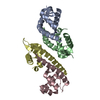
| Title | Crystal Structure of the Acinetobacter baumannii LysR family regulator AceR effector-binding domain with Spermidine | ||||||
 Components Components | Bacterial regulatory helix-turn-helix protein, lysR family protein | ||||||
 Keywords Keywords | TRANSCRIPTION / LysR family regulator / effector-binding domain | ||||||
| Function / homology |  Function and homology information Function and homology informationtranscription cis-regulatory region binding / DNA-binding transcription factor activity Similarity search - Function | ||||||
| Biological species |  Acinetobacter baumannii (bacteria) Acinetobacter baumannii (bacteria) | ||||||
| Method |  X-RAY DIFFRACTION / SYNCHROTRON / MOLECULAR REPLACEMENT / Resolution: 1.97 Å X-RAY DIFFRACTION / SYNCHROTRON / MOLECULAR REPLACEMENT / Resolution: 1.97 Å | ||||||
 Authors Authors | Ma, J.M. / Ge, H.H. | ||||||
| Funding support |  China, 1items China, 1items
| ||||||
 Citation Citation | Journal: To Be Published Title: Crystal Structure of Acinetobacter baumannii LysR family regulator AceR effector binding domain (apo) Authors: Ma, J.M. / Ge, H.H. | ||||||
| History |
|
- Structure visualization
| Structure viewer | Molecule: MolmilJmol/JSmol |
|---|
- Downloads & links
-Download
| PDBx/mmCIF format | 8j4o.cif.gz | 171.2 KB | Display | PDBx/mmCIF format |
|---|---|---|---|---|
| PDB format | pdb8j4o.ent.gz | 132.4 KB | Display | PDB format |
| PDBx/mmJSON format | 8j4o.json.gz | Tree view | PDBx/mmJSON format | |
| Others |  Other downloads Other downloads |
-Validation report
| Arichive directory | https://data.pdbj.org/pub/pdb/validation_reports/j4/8j4oftp://data.pdbj.org/pub/pdb/validation_reports/j4/8j4o | HTTPS FTP |
|---|
-Related structure data


-Links
 PDBj
PDBj
- Assembly
| Deposited unit | 
| ||||||||
|---|---|---|---|---|---|---|---|---|---|
| 1 |
| ||||||||
| Unit cell |
|
-Components
| #1: Protein | Mass: 34169.141 Da / Num. of mol.: 2 / Fragment: effector-binding domain Source method: isolated from a genetically manipulated source Source: (gene. exp.) Acinetobacter baumannii (bacteria) / Gene: cynR_1 / Production host: #2: Chemical | ChemComp-SPD / |   Mass: 145.246 Da / Num. of mol.: 1 / Source method: obtained synthetically / Formula: C7H19N3 / Feature type: SUBJECT OF INVESTIGATION Mass: 145.246 Da / Num. of mol.: 1 / Source method: obtained synthetically / Formula: C7H19N3 / Feature type: SUBJECT OF INVESTIGATION#3: Water | ChemComp-HOH / |  Mass: 18.015 Da / Num. of mol.: 260 / Source method: isolated from a natural source / Formula: H2O Mass: 18.015 Da / Num. of mol.: 260 / Source method: isolated from a natural source / Formula: H2OHas ligand of interest | Y | Has protein modification | N | |
|---|
-Experimental details
-Experiment
| Experiment | Method: X-RAY DIFFRACTION / Number of used crystals: 1 |
|---|
- Sample preparation
| Crystal | Density Matthews: 1.58 Å3/Da / Density % sol: 22.3 % |
|---|---|
| Crystal grow | Temperature: 290 K / Method: vapor diffusion / Details: 1.5 M Ammonium sulfate, 0.1 M Tris-HCl pH8.5 |
-Data collection
| Diffraction | Mean temperature: 80 K / Serial crystal experiment: N |
|---|---|
| Diffraction source | Source: SYNCHROTRON / Site: SSRF / Beamline: BL18U1 / Wavelength: 0.9792 Å |
| Detector | Type: DECTRIS PILATUS3 6M / Detector: PIXEL / Date: Dec 30, 2021 |
| Radiation | Protocol: SINGLE WAVELENGTH / Monochromatic (M) / Laue (L): M / Scattering type: x-ray |
| Radiation wavelength | Wavelength: 0.9792 Å / Relative weight: 1 |
| Reflection | Resolution: 1.966→61.108 Å / Num. obs: 29903 / % possible obs: 94.6 % / Redundancy: 7.2 % / CC1/2: 0.998 / Rmerge(I) obs: 0.117 / Rpim(I) all: 0.046 / Rrim(I) all: 0.126 / Net I/σ(I): 13.2 |
| Reflection shell | Resolution: 1.966→2 Å / Rmerge(I) obs: 1.308 / Mean I/σ(I) obs: 2 / Num. unique obs: 1561 / CC1/2: 0.688 / Rpim(I) all: 0.522 / Rrim(I) all: 1.411 / % possible all: 100 |
- Processing
| Software |
| |||||||||||||||||||||||||||||||||||||||||||||||||||||||||||||||||||||||||||||
|---|---|---|---|---|---|---|---|---|---|---|---|---|---|---|---|---|---|---|---|---|---|---|---|---|---|---|---|---|---|---|---|---|---|---|---|---|---|---|---|---|---|---|---|---|---|---|---|---|---|---|---|---|---|---|---|---|---|---|---|---|---|---|---|---|---|---|---|---|---|---|---|---|---|---|---|---|---|---|
| Refinement | Method to determine structure: MOLECULAR REPLACEMENT Starting model: AlphaFold Resolution: 1.97→55.85 Å / SU ML: 0.23 / Cross valid method: FREE R-VALUE / σ(F): 1.34 / Phase error: 23.92 / Stereochemistry target values: ML
| |||||||||||||||||||||||||||||||||||||||||||||||||||||||||||||||||||||||||||||
| Solvent computation | Shrinkage radii: 0.9 Å / VDW probe radii: 1.11 Å / Solvent model: FLAT BULK SOLVENT MODEL | |||||||||||||||||||||||||||||||||||||||||||||||||||||||||||||||||||||||||||||
| Refinement step | Cycle: LAST / Resolution: 1.97→55.85 Å
| |||||||||||||||||||||||||||||||||||||||||||||||||||||||||||||||||||||||||||||
| Refine LS restraints |
| |||||||||||||||||||||||||||||||||||||||||||||||||||||||||||||||||||||||||||||
| LS refinement shell |
| |||||||||||||||||||||||||||||||||||||||||||||||||||||||||||||||||||||||||||||
| Refinement TLS params. | Method: refined / Origin x: 22.9075 Å / Origin y: 20.7777 Å / Origin z: 2.222 Å
| |||||||||||||||||||||||||||||||||||||||||||||||||||||||||||||||||||||||||||||
| Refinement TLS group | Selection details: all |
