 Movie
Movie Controller


+ Open data

- Basic information
| Entry | Database: PDB / ID: 8j44 | ||||||
|---|---|---|---|---|---|---|---|
| Title | Reductive Aminase RA34-WT | ||||||
 Components Components | 6-phosphogluconate dehydrogenase NAD-binding protein | ||||||
 Keywords Keywords | OXIDOREDUCTASE / Reductive Aminase / keto ester / oxidoreductases | ||||||
| Function / homology |  Function and homology information Function and homology informationtranscription elongation-coupled chromatin remodeling / nucleosome binding / NADP binding / oxidoreductase activity / chromatin / DNA binding Similarity search - Function | ||||||
| Biological species |  Streptomyces viridochromogenes (bacteria) Streptomyces viridochromogenes (bacteria) | ||||||
| Method |  X-RAY DIFFRACTION / MOLECULAR REPLACEMENT / Resolution: 2.25 Å X-RAY DIFFRACTION / MOLECULAR REPLACEMENT / Resolution: 2.25 Å | ||||||
 Authors Authors | Zheng, X.Y. / Xu, G.C. / Ni, Y. | ||||||
| Funding support |  China, 1items China, 1items
| ||||||
 Citation Citation | Journal: To Be Published Title: Dynamic kinetic reductive resolution of cyclic keto esters by newly identified stereo complementary reductive aminases. Authors: Zheng, X.Y. / Xu, G.C. / Ni, Y. | ||||||
| History |
|
- Structure visualization
| Structure viewer | Molecule: MolmilJmol/JSmol |
|---|
- Downloads & links
-Download
| PDBx/mmCIF format | 8j44.cif.gz | 123.2 KB | Display | PDBx/mmCIF format |
|---|---|---|---|---|
| PDB format | pdb8j44.ent.gz | 94.4 KB | Display | PDB format |
| PDBx/mmJSON format | 8j44.json.gz | Tree view | PDBx/mmJSON format | |
| Others |  Other downloads Other downloads |
-Validation report
| Summary document | 8j44_validation.pdf.gz | 1.1 MB | Display | wwPDB validaton report |
|---|---|---|---|---|
| Full document | 8j44_full_validation.pdf.gz | 1.1 MB | Display | |
| Data in XML | 8j44_validation.xml.gz | 24 KB | Display | |
| Data in CIF | 8j44_validation.cif.gz | 33.2 KB | Display | |
| Arichive directory | https://data.pdbj.org/pub/pdb/validation_reports/j4/8j44ftp://data.pdbj.org/pub/pdb/validation_reports/j4/8j44 | HTTPS FTP |
-Related structure data

-Links
 PDBj
PDBj
- Assembly
| Deposited unit | 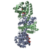
| ||||||||
|---|---|---|---|---|---|---|---|---|---|
| 1 |
| ||||||||
| Unit cell |
|
-Components
| #1: Protein | Mass: 30799.824 Da / Num. of mol.: 2 Source method: isolated from a genetically manipulated source Source: (gene. exp.) Streptomyces viridochromogenes (strain DSM 40736 / JCM 4977 / BCRC 1201 / Tue 494) (bacteria)Gene: SSQG_04344 / Production host: #2: Chemical |   Mass: 743.405 Da / Num. of mol.: 2 / Source method: obtained synthetically / Formula: C21H28N7O17P3 / Feature type: SUBJECT OF INVESTIGATION Mass: 743.405 Da / Num. of mol.: 2 / Source method: obtained synthetically / Formula: C21H28N7O17P3 / Feature type: SUBJECT OF INVESTIGATION#3: Water | ChemComp-HOH / |  Mass: 18.015 Da / Num. of mol.: 150 / Source method: isolated from a natural source / Formula: H2O Mass: 18.015 Da / Num. of mol.: 150 / Source method: isolated from a natural source / Formula: H2OHas ligand of interest | Y | |
|---|
-Experimental details
-Experiment
| Experiment | Method: X-RAY DIFFRACTION / Number of used crystals: 1 |
|---|
- Sample preparation
| Crystal | Density Matthews: 2.61 Å3/Da / Density % sol: 52.88 % |
|---|---|
| Crystal grow | Temperature: 289.2 K / Method: vapor diffusion, sitting drop / pH: 7.4 / Details: Tacsimate, MES monohydrate, PEG 4000 |
-Data collection
| Diffraction | Mean temperature: 150 K / Serial crystal experiment: N |
|---|---|
| Diffraction source | Source: SEALED TUBE / Type: BRUKER D8 QUEST / Wavelength: 1.54178 Å |
| Detector | Type: Bruker PHOTON II / Detector: PIXEL / Date: Nov 25, 2022 / Details: multilayer |
| Radiation | Protocol: SINGLE WAVELENGTH / Monochromatic (M) / Laue (L): M / Scattering type: x-ray |
| Radiation wavelength | Wavelength: 1.54178 Å / Relative weight: 1 |
| Reflection | Resolution: 2.25→23.25 Å / Num. obs: 30171 / % possible obs: 99.1 % / Redundancy: 4.2 % / Rpim(I) all: 0.047 / Net I/σ(I): 12.9 |
| Reflection shell | Resolution: 2.25→2.32 Å / Num. unique obs: 2558 / Rpim(I) all: 0.266 |
- Processing
| Software |
| ||||||||||||||||||||||||||||||||||||||||||||||||||||||||||||||||||||||||||||||||||||||||||||||||||||||||||||||||||||||||||||||||||||||||||||||||||||||||||||||||||||||||||||||||||||||
|---|---|---|---|---|---|---|---|---|---|---|---|---|---|---|---|---|---|---|---|---|---|---|---|---|---|---|---|---|---|---|---|---|---|---|---|---|---|---|---|---|---|---|---|---|---|---|---|---|---|---|---|---|---|---|---|---|---|---|---|---|---|---|---|---|---|---|---|---|---|---|---|---|---|---|---|---|---|---|---|---|---|---|---|---|---|---|---|---|---|---|---|---|---|---|---|---|---|---|---|---|---|---|---|---|---|---|---|---|---|---|---|---|---|---|---|---|---|---|---|---|---|---|---|---|---|---|---|---|---|---|---|---|---|---|---|---|---|---|---|---|---|---|---|---|---|---|---|---|---|---|---|---|---|---|---|---|---|---|---|---|---|---|---|---|---|---|---|---|---|---|---|---|---|---|---|---|---|---|---|---|---|---|---|
| Refinement | Method to determine structure: MOLECULAR REPLACEMENT / Resolution: 2.25→23.25 Å / Cor.coef. Fo:Fc: 0.948 / Cor.coef. Fo:Fc free: 0.929 / SU B: 6.759 / SU ML: 0.161 / Cross valid method: THROUGHOUT / ESU R: 0.292 / ESU R Free: 0.209 / Stereochemistry target values: MAXIMUM LIKELIHOOD / Details: HYDROGENS HAVE BEEN ADDED IN THE RIDING POSITIONS
| ||||||||||||||||||||||||||||||||||||||||||||||||||||||||||||||||||||||||||||||||||||||||||||||||||||||||||||||||||||||||||||||||||||||||||||||||||||||||||||||||||||||||||||||||||||||
| Solvent computation | Ion probe radii: 0.8 Å / Shrinkage radii: 0.8 Å / VDW probe radii: 1.2 Å / Solvent model: MASK | ||||||||||||||||||||||||||||||||||||||||||||||||||||||||||||||||||||||||||||||||||||||||||||||||||||||||||||||||||||||||||||||||||||||||||||||||||||||||||||||||||||||||||||||||||||||
| Displacement parameters | Biso mean: 31.734 Å2
| ||||||||||||||||||||||||||||||||||||||||||||||||||||||||||||||||||||||||||||||||||||||||||||||||||||||||||||||||||||||||||||||||||||||||||||||||||||||||||||||||||||||||||||||||||||||
| Refinement step | Cycle: 1 / Resolution: 2.25→23.25 Å
| ||||||||||||||||||||||||||||||||||||||||||||||||||||||||||||||||||||||||||||||||||||||||||||||||||||||||||||||||||||||||||||||||||||||||||||||||||||||||||||||||||||||||||||||||||||||
| Refine LS restraints |
|