 Movie
Movie Controller

[English] 日本語
 Yorodumi
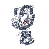
- PDB-8j3y: Crystal structure of CBM6E E168Q in complex with oligosaccharides -
+ Open data

- Basic information
| Entry | Database: PDB / ID: 8j3y | ||||||
|---|---|---|---|---|---|---|---|
| Title | Crystal structure of CBM6E E168Q in complex with oligosaccharides | ||||||
 Components Components | Putative polysaccharide-binding protein | ||||||
 Keywords Keywords | HYDROLASE / glycoside hydrolase | ||||||
| Function / homology |  Function and homology information Function and homology informationfungal-type cell wall polysaccharide metabolic process / carbohydrate binding / metal ion binding Similarity search - Function | ||||||
| Biological species |  Saccharophagus degradans (bacteria) Saccharophagus degradans (bacteria) | ||||||
| Method |  X-RAY DIFFRACTION / SYNCHROTRON / MOLECULAR REPLACEMENT / Resolution: 1.28 Å X-RAY DIFFRACTION / SYNCHROTRON / MOLECULAR REPLACEMENT / Resolution: 1.28 Å | ||||||
 Authors Authors | He, C. / Li, F. | ||||||
| Funding support |  China, 1items China, 1items
| ||||||
 Citation Citation | Journal: Biotechnol Biofuels Bioprod / Year: 2024 Title: Structural insights into curdlan degradation via a glycoside hydrolase containing a disruptive carbohydrate-binding module. Authors: Lv, T. / Feng, J. / Jia, X. / Wang, C. / Li, F. / Peng, H. / Xiao, Y. / Liu, L. / He, C. | ||||||
| History |
|
- Structure visualization
| Structure viewer | Molecule: MolmilJmol/JSmol |
|---|
- Downloads & links
-Download
| PDBx/mmCIF format | 8j3y.cif.gz | 209.9 KB | Display | PDBx/mmCIF format |
|---|---|---|---|---|
| PDB format | pdb8j3y.ent.gz | 162 KB | Display | PDB format |
| PDBx/mmJSON format | 8j3y.json.gz | Tree view | PDBx/mmJSON format | |
| Others |  Other downloads Other downloads |
-Validation report
| Summary document | 8j3y_validation.pdf.gz | 1.8 MB | Display | wwPDB validaton report |
|---|---|---|---|---|
| Full document | 8j3y_full_validation.pdf.gz | 1.8 MB | Display | |
| Data in XML | 8j3y_validation.xml.gz | 43.4 KB | Display | |
| Data in CIF | 8j3y_validation.cif.gz | 68.8 KB | Display | |
| Arichive directory | https://data.pdbj.org/pub/pdb/validation_reports/j3/8j3yftp://data.pdbj.org/pub/pdb/validation_reports/j3/8j3y | HTTPS FTP |
-Related structure data
-Links
 PDBj
PDBj


- Assembly
| Deposited unit | 
| ||||||||
|---|---|---|---|---|---|---|---|---|---|
| 1 | 
| ||||||||
| 2 | 
| ||||||||
| Unit cell |
|
-Components
-Protein , 1 types, 2 molecules AB
| #1: Protein | Mass: 44554.020 Da / Num. of mol.: 2 / Mutation: E168Q Source method: isolated from a genetically manipulated source Source: (gene. exp.) Saccharophagus degradans (strain 2-40 / ATCC 43961 / DSM 17024) (bacteria)Gene: cbm6E, Sde_1445 / Production host: |
|---|
-Sugars , 3 types, 5 molecules 
| #2: Polysaccharide | beta-D-glucopyranose-(1-3)-beta-D-glucopyranose-(1-3)-beta-D-glucopyranose-(1-3)-beta-D- ...beta-D-glucopyranose-(1-3)-beta-D-glucopyranose-(1-3)-beta-D-glucopyranose-(1-3)-beta-D-glucopyranose-(1-3)-beta-D-glucopyranose Source method: isolated from a genetically manipulated source | ||
|---|---|---|---|
| #3: Polysaccharide | Source method: isolated from a genetically manipulated source #5: Sugar | ChemComp-BGC / |  Type: D-saccharide, beta linking / Mass: 180.156 Da / Num. of mol.: 1 / Source method: obtained synthetically / Formula: C6H12O6 / Feature type: SUBJECT OF INVESTIGATION Type: D-saccharide, beta linking / Mass: 180.156 Da / Num. of mol.: 1 / Source method: obtained synthetically / Formula: C6H12O6 / Feature type: SUBJECT OF INVESTIGATION |
-Non-polymers , 3 types, 1337 molecules 


| #4: Chemical |  Mass: 92.094 Da / Num. of mol.: 3 / Source method: obtained synthetically / Formula: C3H8O3 Mass: 92.094 Da / Num. of mol.: 3 / Source method: obtained synthetically / Formula: C3H8O3#6: Chemical |  Mass: 24.305 Da / Num. of mol.: 2 / Source method: obtained synthetically / Formula: Mg Mass: 24.305 Da / Num. of mol.: 2 / Source method: obtained synthetically / Formula: Mg#7: Water | ChemComp-HOH / | Mass: 18.015 Da / Num. of mol.: 1332 / Source method: isolated from a natural source / Formula: H2O |
|---|
-Details
| Has ligand of interest | Y |
|---|---|
| Has protein modification | Y |
-Experimental details
-Experiment
| Experiment | Method: X-RAY DIFFRACTION / Number of used crystals: 1 |
|---|
- Sample preparation
| Crystal | Density Matthews: 2.39 Å3/Da / Density % sol: 48.62 % |
|---|---|
| Crystal grow | Temperature: 283 K / Method: vapor diffusion, sitting drop Details: 0.2 M magnesium chloride, 0.1 M bis-tris, pH 6.5, 25% w/v polyethylene glycol 3,350 |
-Data collection
| Diffraction | Mean temperature: 100 K / Serial crystal experiment: N | ||||||||||||||||||||||||||||||||||||||||||||||||||||||||||||||||||||||||||||||||||||||||||||||||||||||||||||||||||||||||||||||||||||||||||||||||||||||||||||||||||||||||||||||||||||||||||||||||||||||||||||||||||
|---|---|---|---|---|---|---|---|---|---|---|---|---|---|---|---|---|---|---|---|---|---|---|---|---|---|---|---|---|---|---|---|---|---|---|---|---|---|---|---|---|---|---|---|---|---|---|---|---|---|---|---|---|---|---|---|---|---|---|---|---|---|---|---|---|---|---|---|---|---|---|---|---|---|---|---|---|---|---|---|---|---|---|---|---|---|---|---|---|---|---|---|---|---|---|---|---|---|---|---|---|---|---|---|---|---|---|---|---|---|---|---|---|---|---|---|---|---|---|---|---|---|---|---|---|---|---|---|---|---|---|---|---|---|---|---|---|---|---|---|---|---|---|---|---|---|---|---|---|---|---|---|---|---|---|---|---|---|---|---|---|---|---|---|---|---|---|---|---|---|---|---|---|---|---|---|---|---|---|---|---|---|---|---|---|---|---|---|---|---|---|---|---|---|---|---|---|---|---|---|---|---|---|---|---|---|---|---|---|---|---|---|
| Diffraction source | Source: SYNCHROTRON / Site: SSRF / Beamline: BL18U1 / Wavelength: 0.9792 Å | ||||||||||||||||||||||||||||||||||||||||||||||||||||||||||||||||||||||||||||||||||||||||||||||||||||||||||||||||||||||||||||||||||||||||||||||||||||||||||||||||||||||||||||||||||||||||||||||||||||||||||||||||||
| Detector | Type: DECTRIS PILATUS3 6M / Detector: PIXEL / Date: Dec 20, 2020 | ||||||||||||||||||||||||||||||||||||||||||||||||||||||||||||||||||||||||||||||||||||||||||||||||||||||||||||||||||||||||||||||||||||||||||||||||||||||||||||||||||||||||||||||||||||||||||||||||||||||||||||||||||
| Radiation | Protocol: SINGLE WAVELENGTH / Monochromatic (M) / Laue (L): M / Scattering type: x-ray | ||||||||||||||||||||||||||||||||||||||||||||||||||||||||||||||||||||||||||||||||||||||||||||||||||||||||||||||||||||||||||||||||||||||||||||||||||||||||||||||||||||||||||||||||||||||||||||||||||||||||||||||||||
| Radiation wavelength | Wavelength: 0.9792 Å / Relative weight: 1 | ||||||||||||||||||||||||||||||||||||||||||||||||||||||||||||||||||||||||||||||||||||||||||||||||||||||||||||||||||||||||||||||||||||||||||||||||||||||||||||||||||||||||||||||||||||||||||||||||||||||||||||||||||
| Reflection | Resolution: 1.28→40 Å / Num. obs: 215191 / % possible obs: 100 % / Redundancy: 6.6 % / CC1/2: 0.997 / CC star: 0.999 / Rmerge(I) obs: 0.068 / Rpim(I) all: 0.028 / Rrim(I) all: 0.073 / Χ2: 0.91 / Net I/σ(I): 6 / Num. measured all: 1428606 | ||||||||||||||||||||||||||||||||||||||||||||||||||||||||||||||||||||||||||||||||||||||||||||||||||||||||||||||||||||||||||||||||||||||||||||||||||||||||||||||||||||||||||||||||||||||||||||||||||||||||||||||||||
| Reflection shell | Diffraction-ID: 1
|
- Processing
| Software |
| |||||||||||||||||||||||||||||||||||||||||||||||||||||||||||||||||||||||||||||||||||||||||||||||||||||||||||||||||||||||||||||||||||||||||||||||||||||||||||||||||||||||||||||||||||||||||||||||||||||||||||||||||||||||||
|---|---|---|---|---|---|---|---|---|---|---|---|---|---|---|---|---|---|---|---|---|---|---|---|---|---|---|---|---|---|---|---|---|---|---|---|---|---|---|---|---|---|---|---|---|---|---|---|---|---|---|---|---|---|---|---|---|---|---|---|---|---|---|---|---|---|---|---|---|---|---|---|---|---|---|---|---|---|---|---|---|---|---|---|---|---|---|---|---|---|---|---|---|---|---|---|---|---|---|---|---|---|---|---|---|---|---|---|---|---|---|---|---|---|---|---|---|---|---|---|---|---|---|---|---|---|---|---|---|---|---|---|---|---|---|---|---|---|---|---|---|---|---|---|---|---|---|---|---|---|---|---|---|---|---|---|---|---|---|---|---|---|---|---|---|---|---|---|---|---|---|---|---|---|---|---|---|---|---|---|---|---|---|---|---|---|---|---|---|---|---|---|---|---|---|---|---|---|---|---|---|---|---|---|---|---|---|---|---|---|---|---|---|---|---|---|---|---|---|
| Refinement | Method to determine structure: MOLECULAR REPLACEMENT / Resolution: 1.28→21.15 Å / SU ML: 0.11 / Cross valid method: FREE R-VALUE / σ(F): 1.36 / Phase error: 16.42 / Stereochemistry target values: ML
| |||||||||||||||||||||||||||||||||||||||||||||||||||||||||||||||||||||||||||||||||||||||||||||||||||||||||||||||||||||||||||||||||||||||||||||||||||||||||||||||||||||||||||||||||||||||||||||||||||||||||||||||||||||||||
| Solvent computation | Shrinkage radii: 0.9 Å / VDW probe radii: 1.11 Å / Solvent model: FLAT BULK SOLVENT MODEL | |||||||||||||||||||||||||||||||||||||||||||||||||||||||||||||||||||||||||||||||||||||||||||||||||||||||||||||||||||||||||||||||||||||||||||||||||||||||||||||||||||||||||||||||||||||||||||||||||||||||||||||||||||||||||
| Refinement step | Cycle: LAST / Resolution: 1.28→21.15 Å
| |||||||||||||||||||||||||||||||||||||||||||||||||||||||||||||||||||||||||||||||||||||||||||||||||||||||||||||||||||||||||||||||||||||||||||||||||||||||||||||||||||||||||||||||||||||||||||||||||||||||||||||||||||||||||
| Refine LS restraints |
| |||||||||||||||||||||||||||||||||||||||||||||||||||||||||||||||||||||||||||||||||||||||||||||||||||||||||||||||||||||||||||||||||||||||||||||||||||||||||||||||||||||||||||||||||||||||||||||||||||||||||||||||||||||||||
| LS refinement shell |
|
